Review
. 2018 Aug:51:177-186.
doi: 10.1016/j.sbi.2018.07.003. Epub 2018 Jul 23.

Microscopic view of lipids and their diverse biological functions

Affiliations
Review

Microscopic view of lipids and their diverse biological functions

Po-Chao Wen et al. Curr Opin Struct Biol. 2018 Aug.

Abstract

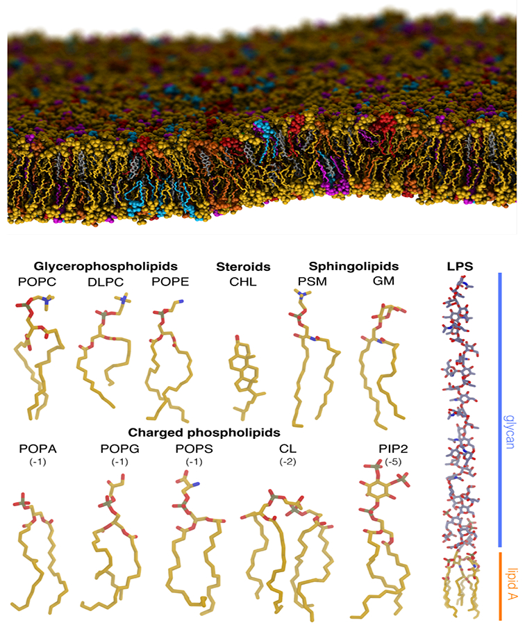
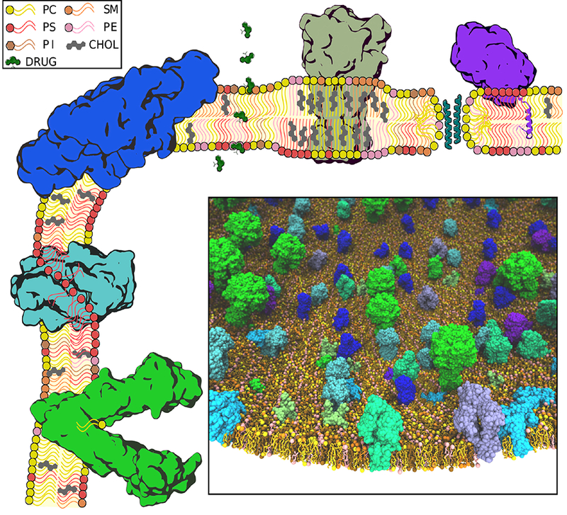
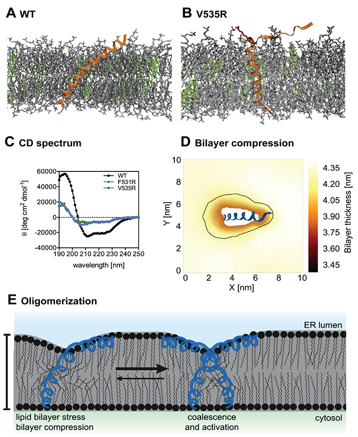
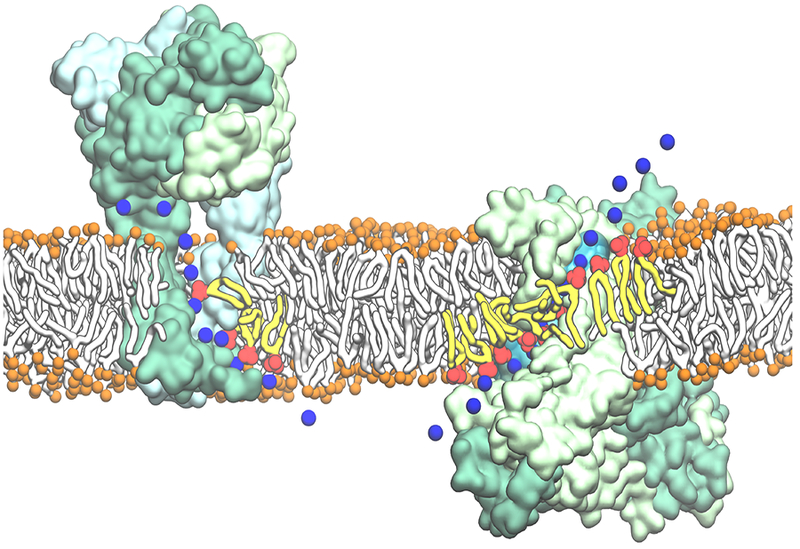
Biological membranes and their diverse lipid constituents play key roles in a broad spectrum of cellular and physiological processes. Characterization of membrane-associated phenomena at a microscopic level is therefore essential to our fundamental understanding of such processes. Due to the semi-fluid and dynamic nature of lipid bilayers, and their complex compositions, detailed characterization of biological membranes at an atomic scale has been refractory to experimental approaches. Computational modeling and simulation offer a highly complementary toolset with sufficient spatial and temporal resolutions to fill this gap. Here, we review recent molecular dynamics studies focusing on the diversity of lipid composition of biological membranes, or aiming at the characterization of lipid-protein interaction, with the overall goal of dissecting how lipids impact biological roles of the cellular membranes.


Figures

Figure 1:
Figure 1:
Heterogeneous lipid composition of biological membranes. (Top) An atomistic MD simulation system of a membrane with a complex, heterogeneous lipid composition. Seven different lipids, including cholesterol, cardiolipin, and a variety of phospholipids, are shown in different colors. Spontaneous curvature of the membrane arising from thermal fluctuations in the simulation can be observed. (Bottom) Structures of some exemplary lipids highlighting a variety of important features (e.g., head group charge and size, tail length and saturation, etc.) associated with lipids. Phospholipids and cholesterol (CHL) are major constituents of cellular membranes. Sphingolipids are common signaling lipids, cardiolipins (CL) are essential mitochondrial lipids, and lipopolysaccharides (LPS) are vital bacterial lipids of the outer membrane.
Figure 2:
Figure 2:
Diverse membrane-associated proteins and various modes of lipid interactions. Various lipid constituents and other small molecules (e.g., drugs) primarily partitioning in the membrane are drawn schematically (see inset key). Exemplary protein components with demonstrated significant interaction with lipids/membrane are drawn using a schematic-looking image based on their actual structure: green: an ABC transporter, bound to a lipid to be transported; cyan: scramblase mediating lipids traversing the two leaflets; blue: envelope and membrane proteins from dengue virus inducing a positive membrane curvature; olive: P2× receptor partitioned in a cholesterol-rich lipid raft, with an increased thickness and higher lipid order; teal: aggregation of antimicrobial peptides resulting in thoroidal pore formation; and, purple: cytochrome P450 anchored to the membrane through an inserted helix. (Inset) A fully atomistic model of a mesoscopic, heterogenous slice of the cellular membrane including various membrane-associated proteins. Several different types of proteins, including both peripheral and integral membrane proteins (shown in different colors) are included in the model. Construction of such complex molecular systems requires methodical placement of proteins and lipids and careful treatment of lipid-protein interfaces to ensure optimal packing.
Figure 3:
Figure 3:
Cholesterol effect on the activation of unfolded protein response. The Ire1-derived sensor peptide and the associated V535R mutant are represented by orange helices in (A) and (B) with the amphipathic helix (AH) highlighted in red. Cholesterols and unsaturated phospholipids (POPC and DOPC) are respectively colored in green and gray. Helicity of the peptides was measured by circular dichroism (CD) spectroscopy (C) and together with MD simulations shows that a conserved structure of the AH is necessary for the sensor peptide to tilt. The presence of cholesterol thickens the membrane, and enhances the membrane compression upon the tilting of the sensor peptide (D). The enhanced bilayer stress induced upon the membrane compression facilitates the oligomerization of Ire1, and promotes the activation of unfolded protein response (E). The images are taken from Halbleib et al [23] with permission from Cell Press.
Figure 4:
Figure 4:
Examples of most direct involvement of phospholipids in biological membrane function, mediated by intimate lipid-protein interactions. Representative snapshots from MD simulations on human P2×3 receptor (Left) [48] and nhTMEM16 scramblase (Right) [49] demonstrating the direct involvement of lipids in ion translocation across the membrane. (Left) Lipids line the cytoplasmic fenestrations of the human P2×3 trimer to allow independent Na+ egress through the lateral cytoplasmic fenestrations during the simulation. (Right) Lipids lining the hydrophilic aqueduct on the surface of the nhTMEM16 scramblase play a structural role in forming a “proteolipidic” pore, which is likely to be used by ions to cross the membrane. The P2×3 trimer and nhTMEM16 dimer are shown in surface representations with each monomer colored in a different shade of green. The lipid headgroups interacting closely with the protein and coordinating permeating ions are shown in red with the tails drawn in yellow; bulk lipids are represented by orange spheres (phosphorus atoms) and white tails. The permeating Na+ ions are shown in time series snapshots (blue spheres). The hydration of the “proteolipidic” pore is illustrated by a transparent cyan surface.

References

    1. Jo S, Cheng X, Lee J, Kim S, Park S-J, Patel DS, Beaven AH, Lee KI, Rui H, Parks S, Lee HS, Roux B, A. D. M. Jr, Klauda JB, Qi Y, Im W, CHARMM-GUI 10 years for biomolecular modeling and simulation, J. Comp. Chem 38 (2017) 1114–1124,

      • CHARMM-GUI is a swiss army knife for constructing membrane simulation systems, yet its functionality reaches far beyond membrane simulations. Some of the functional modules of CHARMM-GUI worth noting within the scope of this article include Membrane Builder, Glycolipid Modeler, LPS Modeler, and Martini Maker.

    1. Bovigny C, Tamò G, Lemmin T, Maïno N, Dal Peraro M, LipidBuilder: a framework to build realistic models for biological membranes, J. Chem. Inf. Model 55 (2015) 2491–2499. - PubMed
    1. Wassenaar TA, Ingólfsson HI, Böckmann RA, Tieleman DP, Marrink SJ, Computational lipidomics with insane: A versatile tool for generating custom membranes for molecular simulations, J. Chem. Theory Comput 11 (2015) 2144–2155,

      • The article describes a versatile method for building membranes, termed insane (INSert membrANE) that uses preset, CG lipid templates to build the membrane, also allowing on-the-fly generation of simple lipid types by specifying the headgroup, linker, and lipid tails, greatly improving our ability to create membranes of any lipid composition.

    1. Stansfeld PJ, Goose JE, Caffrey M, Carpenter EP, Parker JL, Newstead S, Sansom MS, Mem-ProtMD: automated insertion of membrane protein structures into explicit lipid membranes, Structure 23 (2015) 1350–1361,

      • The article describes an automated protocol to model membrane proteins in explicit lipid bilayers, a valuable tool for setting up MD simulations of membrane proteins.

    1. Lyubartsev AP, Rabinovich AL , Force field development for lipid membrane simulations, Biochim. Biophys. Acta Biomembr 1858 (2016) 2483–2497. - PubMed

Publication types