Review
. 2018 Jul 26;9(8):375.
doi: 10.3390/genes9080375.

Blueprints for Biosensors: Design, Limitations, and Applications

Affiliations
Review

Blueprints for Biosensors: Design, Limitations, and Applications

Alexander C Carpenter et al. Genes (Basel). .

Abstract

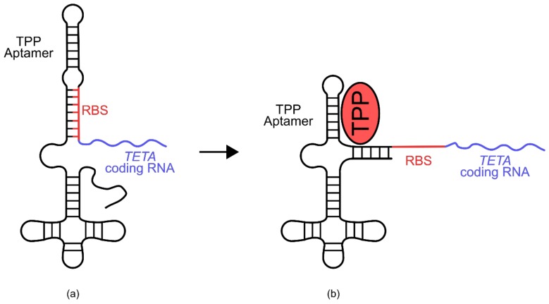
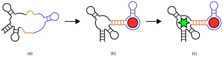
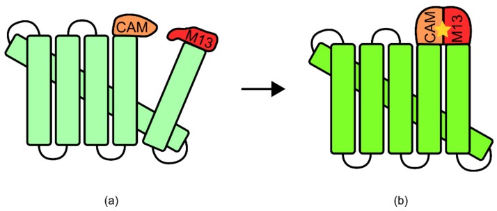
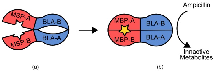
Biosensors are enabling major advances in the field of analytics that are both facilitating and being facilitated by advances in synthetic biology. The ability of biosensors to rapidly and specifically detect a wide range of molecules makes them highly relevant to a range of industrial, medical, ecological, and scientific applications. Approaches to biosensor design are as diverse as their applications, with major biosensor classes including nucleic acids, proteins, and transcription factors. Each of these biosensor types has advantages and limitations based on the intended application, and the parameters that are required for optimal performance. Specifically, the choice of biosensor design must consider factors such as the ligand specificity, sensitivity, dynamic range, functional range, mode of output, time of activation, ease of use, and ease of engineering. This review discusses the rationale for designing the major classes of biosensor in the context of their limitations and assesses their suitability to different areas of biotechnological application.

Keywords: analytics; aptamers; biosensors; high-throughput screening; metabolic engineering; molecular diagnostics; protein switches; synthetic biology.


Conflict of interest statement

The authors declare no conflict of interest. The founding sponsors had no role in the design of the study; in the collection, analyses, or interpretation of data; in the writing of the manuscript, and in the decision to publish the results.

Figures

Figure 1
Figure 1
Transcription factor biosensor configuration. A known transcription factor protein that is regulated by a ligand of interest can be used to target a cognate promoter sequence that is used to initiate transcription of a response gene such as GFP or antibiotic resistance: (a) In the absence of ligand binding to the transcription factor, there is little or no recruitment of RNA polymerase to the promoter site; (b) When ligand is present at a concentration sufficient to bind transcription factor proteins, the transcription factor becomes localized to the promoter; (c) Recruitment of RNA polymerase then allows transcription of the response gene (e.g., GFP or antibiotic resistance).
Figure 2
Figure 2
Butanol biosensor developed for use in E. coli. PBMOR is upstream of the transcriptional regulator gene BMOR. PBMO is upstream of a tetracycline resistance-green fluorescent protein fusion gene (TETA-GFP): (a) under low butanol concentrations, leaky expression driven by PBMOR produces an insignificant amount of BmoR; (b) Increases in butanol concentrations allows coordination of BmoR to PBMOR and PBMO. This allows recruitment of RNA polymerase and the expression of TETA-GFP, producing a cell survival and fluorescence signal. Additionally, by placing PBMOR as the controller of BMOR transcription, this signal is self-amplifying.
Figure 3
Figure 3
Xylose biosensor developed for use in S. cerevisiae. The XYLR gene is under the regulatory control of PHXT7. GFP is under the regulatory control of PGPM1 with an inserted XylR binding site (BS); (a) Under low xylose concentrations, XYLR is constitutively expressed by PHXT7, and binds to the XylR BS, inhibiting the ability of RNA polymerase and other transcriptional machinery from transcribing the downstream GFP gene. (b) Under increased xylose concentrations, the XylR protein binds xylose, disrupting its ability to associating with XylR BS. Free of impediment, this allows PGPM1 to induce the transcription of GFP. Thus, increases in xylose concentrations correlate with an increase in GFP signal.
Figure 4
Figure 4
Isopentenyl-pyrophosphate (IPP) biosensor for use in E. coli. An IPP isomerase (Idi) domain-tether-AraC DNA binding domain fusion protein was expressed under the regulatory control of a constitutive promoter. Whilst an mCherry gene was under the regulatory control of PBAD: (a) Under low IPP concentrations, Idi-linker-AraC DNA binding domains (DBD) fusion protein associated with PBAD and the AraC DBD induced transcription of mCherry; (b) Under increased IPP concentrations, Idi-mediated fusion protein dimerization inhibited the ability of AraC DBD from efficiently inducing transcription of mCherry. Thus, increases in IPP concentration were correlated with decreases in the mCherry signal.
Figure 5
Figure 5
Isopentenyl-pyrophosphate (IPP) biosensor for use in S. cerevisiae. Two fusion proteins were used in the biosensor. The first was IPP isomerase (Idi)-tether-Gal4 activation domain (Gal4 AD). The second was IPP isomerase (Idi)-tether-Gal4 DNA binding domain (Gal4 DBD). Both fusion protein genes were under the regulatory control of constitutive promoters. Also used in this biosensor was a yECitrine gene under the control of PGAL10: (a) Under low IPP concentrations, the fusion proteins were expressed, but had no effect on yECitrine expression; (b) Under increased IPP concentrations, the two fusion proteins are able to dimerize via the Idi domains. Gal4 DBD localized the dimer to PGAL10, while Gal4 AD induced the transcription of yECitrine. Thus, increases in IPP concentration were correlated with increases in the yECitrine signal.
Figure 6
Figure 6
Ochratoxin A biosensor. The biosensor is comprised of a DNA aptamer for Ochratoxin A (a) aptamer with no added SYBR Green or Ochratoxin A; (b) coordinated with SYBR Green (green star); (c) and coordinated with OTA (red oval) and SYBR Green. Coordination of OTA to the aptamer results in decreased binding sites for SYBR Green, and therefore decreased fluorescence. Figure adapted from McKeague, et al. [65].
Figure 7
Figure 7
Thrombin biosensor. Thrombin-binding aptamer immobilized on a gold electrode with covalently added methylene blue group (MB). (a) In the absence of thrombin, the flexible aptamer structure allows the MB group to contact the electrode, donating an electron and generating a signal. (b) Addition of thrombin stabilizes secondary structure, reducing contact of the MB group with the electrode, reducing signal generation. Figure adapted from Xiao, et al. [7].
Figure 8
Figure 8
The systematic evolution of ligands by exponential enrichment (SELEX) process. The SELEX process uses a series of incubation and wash steps to identify single-stranded (ss) DNA or ssRNA molecules with binding affinity to a target ligand. Positive and negative selection steps are employed to avoid binding affinity to structurally similar compounds. Polymerase chain reaction (PCR) is used to amplify positive sequences and enrich them as a proportion of the total population.
Figure 9
Figure 9
Thiamine pyrophosphate (TPP) biosensor. The biosensor is comprised of a TPP-binding aptamer, a TETA-coding RNA, and a modified spacer sequence containing the ribosomal binding site (RBS): (a) Biosensor without TPP. Base pair homology keeps the RBS inaccessible to the ribosome, preventing translation of the TETA-coding RNA; (b) TPP biosensor coordinated to TPP. Coordination allows a more energetically favorable secondary structure to be adopted, which liberates the RBS, allowing translation of the TETA-coding RNA. Under increased TPP concentration conditions, E. coli cells containing this biosensor are able to grow in the presence of the antibiotic tetracycline. Figure adapted from Muranaka, et al. [77].
Figure 10
Figure 10
General Spinach based biosensors. (a) The Spinach aptamer (black) is disrupted with linker sequences (orange) and a ligand binding aptamer (blue), inhibiting its ability to bind the fluorophore 3,5-difluoro-4-hydroxybenzylidene imadazolinone (DFHBI). (b) Coordination of the target ligand (red circle) to the ligand binding aptamer provides sufficient stability to allow the linker regions to assemble. This in turn allows the Spinach aptamer to form its native secondary structure. (c) The reformation of the Spinach aptamer allows the coordination and activation of DFHBI (green star), resulting in ligand dependent fluorescent signal generation. Figure adapted from Paige, et al. [78].
Figure 11
Figure 11
Calcium biosensor. This biosensor is one fusion protein comprised of a cyclically permuted green fluorescent protein (GFP), and calmodulin (CAM), and M13 domains: (a) At low calcium concentrations, the CAM and M13 domains do not coordinate efficiently, disrupting the structure of the cyclically permuted GFP. This disruption reduces the fluorescent output of the protein. (b) Upon binding of calcium (yellow star), the CAM and M13 domains form a more compact and coordinated configuration, allowing the GFP to return to its natural state, increasing fluorescence. Figure adapted from Nagai, et al. [90].
Figure 12
Figure 12
Maltose Biosensor. This biosensor is one fusion protein comprised of a maltose-binding peptide (MBP) and a β-lactamase enzyme (BLA), each of which has been split into two portions (A and B): (a) In the absence of maltose, the MBP-A and MBP-B domains exist in an open hinge conformation, disrupting the structure of the fused BLA-A and BLA-B domains. (b) Upon binding of maltose (star) to the MBP-A and MBP-B domains, this hinge closes, bringing the BLA-A and BLA-B domains back into the correct position. This reconstitutes the enzymatic activity of the BLA protein, allowing cells expressing this biosensor to grow in the presence of β-lactam antibiotics as a signal output.
Figure 13
Figure 13
General pyrroloquinoline quinone glucose dehydrogenase (GDH)-based biosensors. Each portion of a split GDH protein is expressed with a linker to a ligand binding domain (LBD-A or LBD-B): (a) Under conditions of low target ligand, the GDH domains do not interact; (b) the addition of glucose does not restore GDH domain assembly; (c) target ligand (star) binding to both LBD-A and LBD-B brings GDH-A and GDH-B into close proximity, restoring electron transfer ability. Donation of an electron to an electrode acts as the signal output. Figure adapted from Guo, et al. [95].
Figure 14
Figure 14
ARVCF biosensor. The biosensor utilizes the fibronectin type III domain (FN3), human erbin protein domain (PDZ), and the fluorescence resonance energy transfer (FRET) pairs YPet and CyPet. The FN3 and PDZ domains are connected by a semi rigid linker: (a) Under low ARVCF concentrations, this linker keeps these domains separated and brings the YPet and CyPet into close proximity; (b) The binding of ARVCF to the FN3 and PDZ domains provides enough stability to overcome the separating linker rigidity. This pivoting about the linker separates the YPet and CyPet domains, providing a change in FRET energy and generating a change in signal intensity. Figure adapted from Huang and Koide [100].
Figure 15
Figure 15
Rapamycin biosensor built using two separate fusion proteins. The first fusion protein is a rapamycin-binding FRB domain attached via a tether to a hepatitis C virus NS3 serine protease (HCV), which is in turn tethered to a HCV auto-inhibition domain (AI). The second protein is a complementary rapamycin binding FKBP12 domain, tethered to a tobacco vein mottling virus NIa protease (TVMV), which is in turn tethered to a somewhat leaky TVMV auto-inhibition domain (AI): (a) Under conditions of low rapamycin concentration, these proteins do not contact each other; (b) Under increased rapamycin concentrations, the FRB and FKBP domains simultaneously bind rapamycin, bringing the two separate fusion proteins into close proximity; (c) This proximity allows the leaky TVMV protease domain to cleave the tether connecting the HCV protease to its auto-inhibitor; (d) The loss of the auto-inhibitory domain allows the HCV protease to cleave an added fluorogenic peptide with attached quencher, restoring its fluorescence and generating a signal. Figure adapted from Stein and Alexandrov [102].

References

    1. Way J.C., Collins J.J., Keasling J.D., Silver P.A. Integrating biological redesign: Where synthetic biology came from and where it needs to go. Cell. 2014;157:151–161. doi: 10.1016/j.cell.2014.02.039. - DOI - PubMed
    1. Chao R., Mishra S., Si T., Zhao H. Engineering biological systems using automated biofoundries. Metab. Eng. 2017;42:98–108. doi: 10.1016/j.ymben.2017.06.003. - DOI - PMC - PubMed
    1. Hughes R.A., Ellington A.D. Synthetic DNA synthesis and assembly: Putting the synthetic in synthetic biology. Cold Spring Harb. Perspect. Biol. 2017;9 doi: 10.1101/cshperspect.a023812. - DOI - PMC - PubMed
    1. Marcellin E., Nielsen L.K. Advances in analytical tools for high throughput strain engineering. Curr. Opin. Biotechnol. 2018;54:33–40. doi: 10.1016/j.copbio.2018.01.027. - DOI - PubMed
    1. Turner A.P. Biosensors: Sense and sensibility. Chem. Soc. Rev. 2013;42:3184–3196. doi: 10.1039/c3cs35528d. - DOI - PubMed

LinkOut - more resources