. 2018 Jul 26;9(1):2926.
doi: 10.1038/s41467-018-05301-6.

High sensitivity organic inorganic hybrid X-ray detectors with direct transduction and broadband response

Affiliations

High sensitivity organic inorganic hybrid X-ray detectors with direct transduction and broadband response

H M Thirimanne et al. Nat Commun. .

Abstract

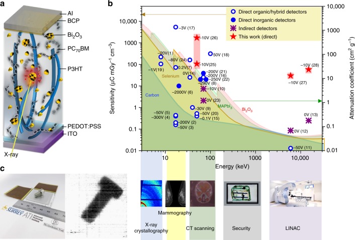
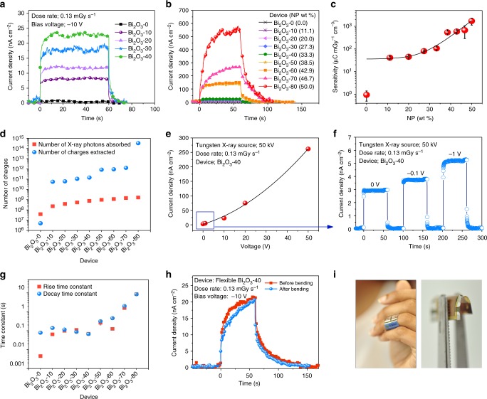
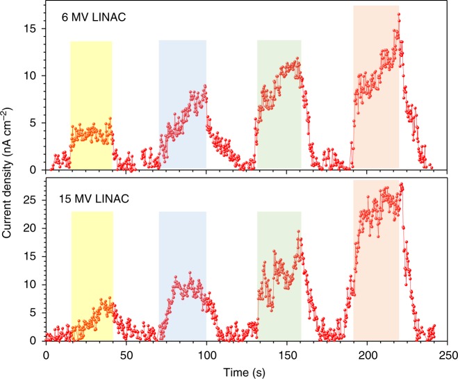
X-ray detectors are critical to healthcare diagnostics, cancer therapy and homeland security, with many potential uses limited by system cost and/or detector dimensions. Current X-ray detector sensitivities are limited by the bulk X-ray attenuation of the materials and consequently necessitate thick crystals (~1 mm-1 cm), resulting in rigid structures, high operational voltages and high cost. Here we present a disruptive, flexible, low cost, broadband, and high sensitivity direct X-ray transduction technology produced by embedding high atomic number bismuth oxide nanoparticles in an organic bulk heterojunction. These hybrid detectors demonstrate sensitivities of 1712 µC mGy-1 cm-3 for "soft" X-rays and ~30 and 58 µC mGy-1 cm-3 under 6 and 15 MV "hard" X-rays generated from a medical linear accelerator; strongly competing with the current solid state detectors, all achieved at low bias voltages (-10 V) and low power, enabling detector operation powered by coin cell batteries.


Conflict of interest statement

The authors declare no competing interests.

Figures

Fig. 1
Fig. 1
X-ray detector overview. a Device schematic structure. b Performance comparison of current solid state X-ray detectors—(1), (2), (3), (4), (5), (7), (9), (11), (14), (15), (16), (17), (18), (19), (20), and (24) are direct detectors, (6), (8), (21), and (22) are inorganic detectors, and (10), (12), (13) and (23) are indirect detectors—with the technology developed in this work—(25) Bi2O3-40, (26) Bi2O3-80, (27) Bi2O3-40 and (28) Bi2O3-40. The operating voltage is given adjacent to each data point. The total attenuation coefficient values of carbon, selenium, methylammonium lead iodide (MAPbI3) and Bi2O3 are given as shaded areas showing the previous limits to detector technology based only on bulk attenuation processes. c An X-ray imager based on the hybrid X-ray detector and 70 kV X-ray image of a bolt taken using the X-ray imager
Fig. 2
Fig. 2
Detector performance under X-ray irradiation. a X-ray photocurrent densities for devices with different Bi2O3 loadings from Bi2O3-0 to Bi2O3-40 and b from Bi2O3-0 to Bi2O3-80. c Averaged sensitivity values for six devices and the error bars represent the range of the sensitivity values. d Comparison between number of X-ray photons absorbed by each device and the number of charges extracted. e The voltage dependence of the Bi2O3-40 device, f X-ray photocurrent response of the Bi2O3-40 detector under 0, −0.1 and −1 V biases. g Rise and decay time constants (the error bars represent the standard error shown with respect to the fitted curves) for detectors with increasing Bi2O3 loadings under −10 V bias. h X-ray photocurrent response before and after bending for a flexible Bi2O3-40 device. i A prototype X-ray detector integrated into a plaster and detector bend radius (0.3 cm)
Fig. 3
Fig. 3
Hard X-ray irradiation. Response of a Bi2O3-40 device under linear accelerator (LINAC) generated 6 and 15 MV X-rays, the consecutive peaks represent the response to dose rates of 114, 227, 340 and 454 µGy s−1
Fig. 4
Fig. 4
X-ray detector structural characterisation. a Grazing incidence wide angle X-ray scattering images of Bi2O3-0, Bi2O3-20, Bi2O3-40, Bi2O3-60 and Bi2O3-80. Here, the position of the poly(3-hexylthiophene-2,5-diyl) (P3HT), [6,6]-Phenyl C71 butyric acid methyl ester (PCBM) and bismuth oxide (Bi2O3) resultant peaks are indicated. b Scanning electron microscopy cross sections of Bi2O3-10, Bi2O3-20 and Bi2O3-40 overlaid with energy dispersive X-ray spectroscopy imaging of the Bi2O3 nanoparticles (false coloured for clarity)
Fig. 5
Fig. 5
Charge transport analysis. a Time-of-flight transients of electrons for the Bi2O3-20 device for the applied reverse-bias voltages from 8 to 20 V. b The double logarithmic plot of the time of flight transient presented in a. Electric field dependency of c electron mobility (data have been fitted to show the mobility dependency under the electric field) and d hole mobility of devices from Bi2O3-0 to Bi2O3-80. Here, the error bars represent the range of three measurements carried out under each condition. e The collected electron charge for different devices based on a fit to the Hecht equation
Fig. 6
Fig. 6
X ray scattering by nanoparticles. a Simulated differential cross sections for X-ray scattering from Bi2O3 NPs with a diameter (d) of 40 nm. b The schematic of the inelastic Mie-scattering mechanism and c the mass attenuation coefficient curve of Bi2O3. Here, Ei, Ea and Es are the energy of the incident X-ray photon, energy absorbed and the energy of the scattered photon, respectively. d The comparison of simulated differential cross section and extracted 1D data of experimental grazing incidence small angle X-ray scattering (GI-SAXS) at 8 keV energy for films with d = 20, 40 and 100 nm NPs. (The dotted line in a represents the extracted data at 8 keV.) e X-ray photocurrent response of the polymer diodes containing Bi2O3 NP sizes of 20, 40 and 100 nm and f the current density value with the spread for six detectors

References

    1. Milbrath, B. D. et al. Radiation detector materials: An overview. J. Mater. Res.23, 2561–2581 (2008).
    1. Zhao W, Ji WG, Debrie A, Rowlands JA. Imaging performance of amorphous selenium based flat-panel detectors for digital mammography: Characterization of a small area prototype detector. Med. Phys. 2003;30:254–263. doi: 10.1118/1.1538233. - DOI - PubMed
    1. Mills CA, et al. Direct detection of 6 MV X-rays from a medical linear accelerator using a semiconducting polymer diode. Phys. Med. Biol. 2013;58:4471–4482. doi: 10.1088/0031-9155/58/13/4471. - DOI - PubMed
    1. Podgorsak EB. Radiation oncology physics: a handbook for teachers and students. Br. J. Cancer. 2008;98:1020. doi: 10.1038/sj.bjc.6604224. - DOI
    1. Takahashi T, Watanabe S. Recent progress in CdTe and CdZnTe detectors. IEEE Trans. Nucl. Sci. 2001;48:950–959. doi: 10.1109/23.958705. - DOI

Publication types