. 2018 Jul 26;8(1):11276.
doi: 10.1038/s41598-018-29310-z.

Involvement of median preoptic nucleus and medullary noradrenergic neurons in cardiovascular and sympathetic responses of hemorrhagic rats

Affiliations

Involvement of median preoptic nucleus and medullary noradrenergic neurons in cardiovascular and sympathetic responses of hemorrhagic rats

Lara Marques Naves et al. Sci Rep. .

Abstract

The infusion of hypertonic saline solution (HSS) is known to be beneficial to the treatment of hypovolemic hemorrhage (HH). The central mechanism of HSS-induced cardiovascular and autonomic recovery of animals subjected to HH remains unclear. Hence, the present study evaluated the involvement of median preoptic nucleus (MnPO) and medullary noradrenergic neurons (A1 and A2) in HSS-induced cardiovascular and sympathetic responses in hemorrhagic rats. The wistar rats were subjected to specific lesion of noradrenergic neurons through the nanoinjections of anti-DβH-saporin into caudal ventrolateral medulla (A1 neurons) and nucleus of the solitary tract (A2 neurons). After recovery, mean arterial pressure (MAP) and renal sympathetic nervous activity were recorded. The HH was performed through blood withdrawal until a MAP of 60 mmHg was attained. In sham rats, HSS infusion (3M NaCl) reestablished MAP without change in HH-induced sympathoinhibition. The muscimol (agonist of GABAA receptor) was nanoinjected in MnPO during HH and MnPO inhibition abolished the recovery of MAP and HSS-induced sympathoinhibition. Simultaneous lesions of A1 and A2 abolished MAP restoration and sympathoinhibition after HSS infusion. These results suggest that the recovery of MAP and HSS-induced sympathoinhibition in hemorrhaged rats depend on intact neural projections from A1 and A2 to MnPO.


Conflict of interest statement

The authors declare no competing interests.

Figures

Figure 1
Figure 1
(A) Representative photomicrograph of coronal section of the hypothalamus (40 μm) showing a typical site of nanoinjection in the median preoptic nucleus (MnPO). The arrow indicates the marking of the MnPO with evans blue dye 4%. AC: anterior commissure. The scale bar is equal to 500 µm. (B) Effects of MnPO pharmacological inhibition on changes in Mean arterial pressure (MAP), (C) heart rate (HR) and (D) integral of renal sympathetic nervous activity (∫RSNA) variations in the rats submitted to hypovolemic hemorrhagic in the MnPO Sham (MnPOS; n = 6) and MnPO Inhibition (MnPOI; n = 6) groups. Values were expressed as means ± standard error of the mean (S.E.M). *Different from time 0; different from sham group with p < 0.05. The black bar represents the hemorrhage period. The dashed line represents the HSS infusion (3M; NaCl). The arrow indicates nanoinjection in the MnPO.
Figure 2
Figure 2
(A) Representative photomicrograph of coronal medulla section (40 μm) marked by immunohistochemistry. The arrows indicate the marking of TH positive neurons in the nucleus of the solitary tract (NTS - cluster A2), in caudal ventrolateral medulla (CVLM - cluster A1) and rostrovetrolateral medulla region (RVLM - cluster C1) of the animals receiving nanoinjection of bilateral saporin (A2S) or saporin-anti-DβH (A2L) in NTS. The scale bar is equal to 100 µm. (B) Quantification of the number of TH positive cells in the medullary regions. Mean ± standard error of the mean (S.E.M.) of the number of positive TH cells located in the nucleus of the solitary tract (NTS – clusters A2 and C2), the caudal ventrolateral medulla (CVLM – cluster A1) and rostral vetrolateral medulla region (RVLM - cluster C1) of the animals receiving nanoinjection of bilateral saporin (A2S) or saporin-anti-DβH (A2L) in NTS. Different from sham group with p < 0.05. (C) Effects of A2 catecholaminergic neuron lesions on changes in Mean arterial pressure (MAP), (D) heart rate (HR) and (E) integral of renal sympathetic nervous activity (∫RSNA) variations in the rats submitted to hypovolemic hemorrhagic in the A2 Sham (A2S; n = 6) and A2 lesioned (A2L; n = 6) groups. Values were expressed as means ± standard error of the mean (S.E.M). *Different from time 0; different from sham group with p < 0.05. The black bar represents the hemorrhage period. The dashed line represents HSS infusion (3M; NaCl).
Figure 3
Figure 3
(A) Representative photomicrograph of coronal medulla section (40 μm) marked by immunohistochemistry. The arrows indicate the marking of TH positive neurons in the nucleus of the solitary tract (NTS - cluster A2), in caudal ventrolateral medulla (CVLM - cluster A1) and rostrovetrolateral medulla region (RVLM - cluster C1) of the animals receiving nanoinjection of bilateral saporin (A1S) or saporin-anti-DβH (A1L) in CVLM. The scale bar is equal to 100 µm. (B) Quantification of the number of TH positive cells in the medullary regions. Mean ± standard error of the mean (S.E.M.) of the number of positive TH cells located in the nucleus of the solitary tract (NTS – clusters A2 and C2), the caudal ventrolateral medulla (CVLM – cluster A1) and rostral vetrolateral medulla region (RVLM - cluster C1) of the animals receiving nanoinjection of bilateral saporin (A1S) or saporin-anti-DβH (A1L) in CVLM. different from sham group with p < 0.05. (C) Effects of A1 + A2 catecholaminergic neuron lesions on changes in Mean arterial pressure (MAP), (D) heart rate (HR) and (E) integral of renal sympathetic nervous activity (∫RSNA) variations in the rats submitted to hypovolemic hemorrhagic in the A1 Sham (A1S; n = 6) and A1 lesioned (A1L; n = 6) groups. Values were expressed as means ± standard error of the mean (S.E.M). *Different from time 0; different from sham group with p < 0.05. The black bar represents the hemorrhage period. The dashed line represents HSS infusion (3M; NaCl).
Figure 4
Figure 4
(A) Representative photomicrograph of coronal medulla section (40μm) marked by immunohistochemistry. The arrows indicate the marking of TH positive neurons in the nucleus of the solitary tract (NTS - cluster A2), in caudal ventrolateral medulla (CVLM - cluster A1) and rostral vetrolateral medulla region (RVLM - cluster C1) of the animals receiving nanoinjection of bilateral saporin (A1 + A2S) or saporin-anti-DβH (A1 + A2L) in CVLM and NTS. The scale bar is equal to 100 µm. (B) Quantification of the number of TH positive cells in the medullary regions. Mean ± standard error of the mean (S.E.M.) of the number of positive TH cells located in the nucleus of the solitary tract (NTS – clusters A2 and C2), the caudal ventrolateral medulla (CVLM – cluster A1) and rostral vetrolateral medulla region (RVLM - cluster C1) of the animals receiving nanoinjection of bilateral saporin (A1 + A2S) or saporin-anti-DβH (A1 + A2L) in CVLM and NTS. Different from sham group with p < 0.05. (C) Effects of A1 + A2 catecholaminergic neuron lesions on changes in Mean arterial pressure (MAP), (D) heart rate (HR) and (E) integral of renal sympathetic nervous activity (∫RSNA) variations in the rats submitted to hypovolemic hemorrhagic in the A1 + A2 Sham (A1 + A2S; n = 6) and A1 + A2 lesioned (A1 + A2L n = 6) groups. Values were expressed as means ± standard error of the mean (S.E.M). *Different from time 0; different from sham group with p < 0.05. The black bar represents the hemorrhage period. The dashed line represents HSS infusion (3M; NaCl).
Figure 5
Figure 5
(A) Effects of A1 + A2 catecholaminergic neuron lesions and MnPO inhibition on changes in mean arterial pressure (Δ% MAP), (B) heart rate (Δ% HR) and (C) integral of renal sympathetic nervous activity (Δ% ∫RSNA) variations in the rats submitted to hypovolemic hemorrhagic in the A1 + A2 lesioned (A1 + A2L; n = 6) and MnPO inhibition (MnPOI; n = 6) groups. Values were expressed as means ± standard error of the mean (S.E.M.). *Different from time 0; different from A1 + A2L group with p < 0.05. The black bar represents the hemorrhage period. The dashed line represents HSS infusion (3M; NaCl).
Figure 6
Figure 6
Post-systolic spectrum of (A,B) arterial pressure (AP), (C,D) renal sympathetic nerve activity (SNA) and coherence values between AP and RSNA power spectrum (E,F) in the rats submitted to hypovolemic hemorrhagic in the MnPO Sham (MnPOS; n = 6) and MnPO Inhibition (MnPOI n = 6) groups.
Figure 7
Figure 7
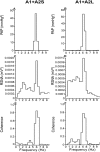
Post-systolic spectrum of (A,B) arterial pressure (AP), (C,D) renal sympathetic nerve activity (SNA) and coherence values between AP and RSNA power spectrum (E,F) in the rats submitted to hypovolemic hemorrhagic in the A1 + A2 Sham (A1 + A2S; n = 6) and A1 + A2 lesioned (A1 + A2L n = 6) groups.
Figure 8
Figure 8
Central pathways involved in the resuscitation of hypertensive hemorrhage promoted by sodium overload. Caudal ventrolateral medulla (CVLM), nucleus of the solitary tract (NTS), magnocellular hypothalamic paraventricular nucleus (mPVN), parvocellular hypothalamic paraventricular nucleus (pPVN), median preoptic nucleus (MnPO), subfornical organ (SFO), organum vasculosum lamina terminalis (OVLT), rostrovetrolateral medulla region (RVLM), oxytocin (OT), vasopressin (VP).

References

    1. Kobayashi L, Costantini TW, Coimbra R. Hypovolemic Shock Resuscitation. Surg. Clin. North Am. 2012;92:1403–1423. doi: 10.1016/j.suc.2012.08.006. - DOI - PubMed
    1. Angele MK, Schneider CP, Chaudry IH. Bench-to-bedside review: latest results in hemorrhagic shock. Crit. Care. 2008;12:218. doi: 10.1186/cc6919. - DOI - PMC - PubMed
    1. Xu D, et al. Sodium hydrosulfide alleviates lung inflammation and cell apoptosis following resuscitated hemorrhagic shock in rats. Acta Pharmacol. Sin. 2013;34:1515–25. doi: 10.1038/aps.2013.96. - DOI - PMC - PubMed
    1. Aletti F, et al. Peptidomic Analysis of Rat Plasma: Proteolysis in Hemorrhagic Shock. Shock. 2016;45:540–54. doi: 10.1097/SHK.0000000000000532. - DOI - PMC - PubMed
    1. Dilmen S, et al. Comparison of warm fluid and cold fluid resuscitation during uncontrolled hemorrhagic shock model in rats. Ulus. Travma Acil Cerrahi Derg. 2017;23:287–293. - PubMed

Publication types

MeSH terms