

Cervical sagittal alignment as a predictor of adjacent-level ossification development
- PMID: 30050320
- PMCID: PMC6055838
- DOI: 10.2147/JPR.S160472
Cervical sagittal alignment as a predictor of adjacent-level ossification development
Abstract
Purpose: To explore the role of cervical sagittal alignment in the occurrence of adjacent-level ossification development (ALOD) in patients who underwent anterior cervical discectomy fusion with self-locking stand-alone polyetheretherketone cage, and the relationship between cervical sagittal alignment and clinical outcomes.
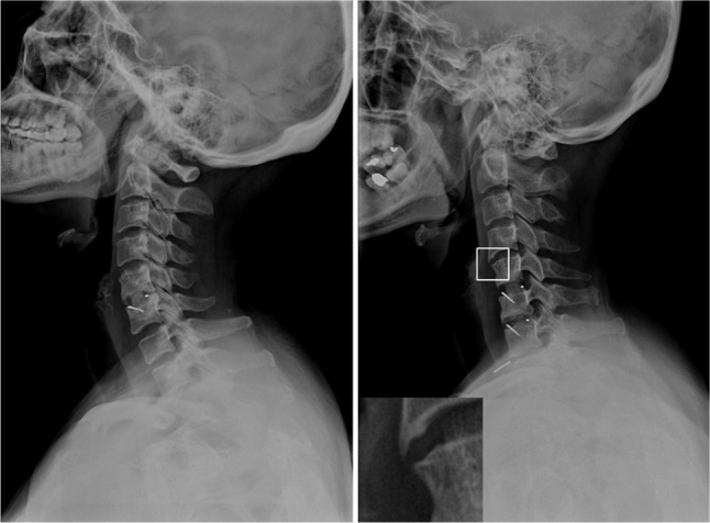
Background: Because of its advantages, anterior cervical plating systems have been used as the classic surgical method in the treatment of patients with cervical disc herniation. However, the proximity (<5 mm) of the plate to the adjacent disc space has proven to be a critical risk factor for ALOD. How cervical sagittal alignment influences the development of ALOD is unknown and its role in ALOD needs clarification.
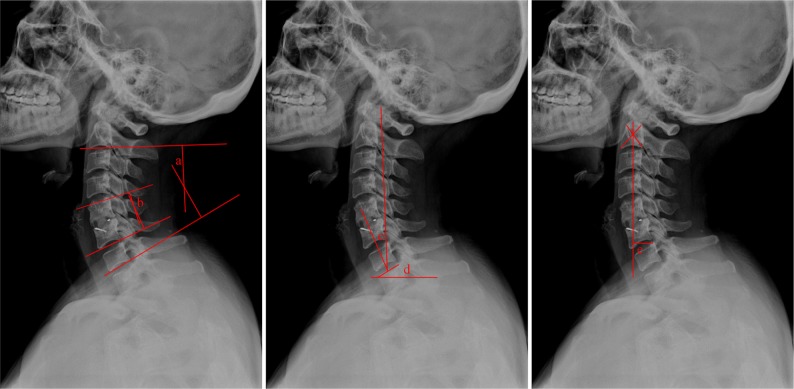
Patients and methods: One hundred and eighteen adults who underwent anterior cervical discectomy fusion with self-locking stand-alone polyetheretherketone cage for cervical radiculopathy or myelopathy between December 2013 and December 2015 were retrospectively recruited. Of these, 15 patients developed ALOD and 103 patients did not, representing two groups for comparison. The cervical sagittal parameters were measured, including C2-C7 Cobb angle (Cobb), fused segment angle, cervical tilt (CT), T1 slope (T1S) and C2-C7 sagittal vertical axis. Clinical outcomes and efficacy were evaluated using a visual analog scale, Japanese Orthopedic Association (JOA) score and neck disability index (NDI) score before and after surgery.
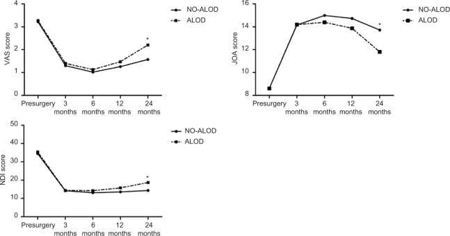
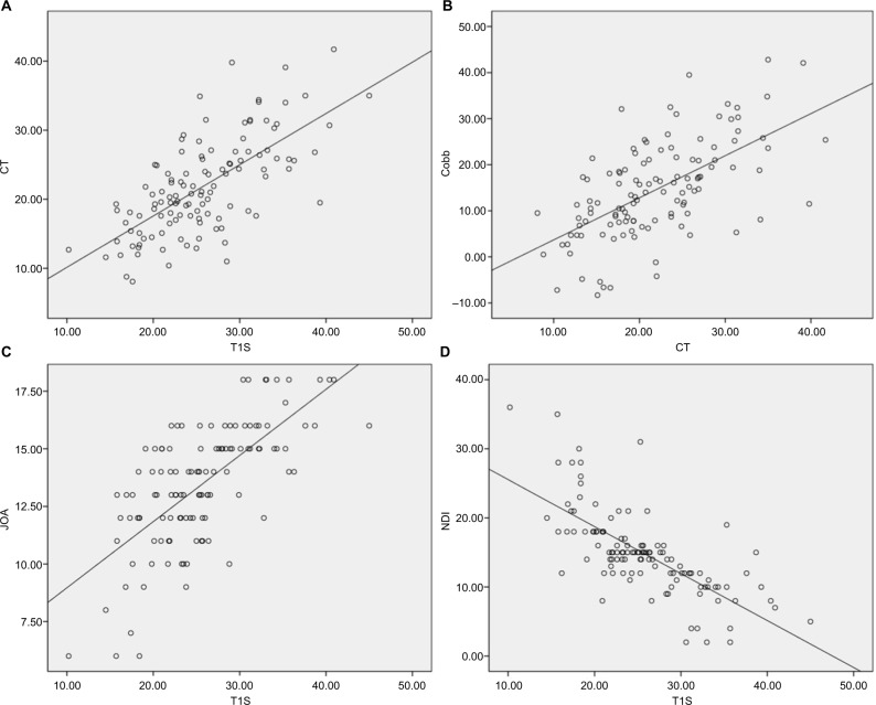
Results: There were no significant differences in patient demographics between the two groups. Cobb value (P<0.05), CT (P<0.05) and T1S (P<0.05) were significantly different between the two groups, while fused segment angle (P>0.05) and C2-C7 sagittal vertical axis (P>0.05) showed no difference. Compared with preoperative scores, improvement was seen in postoperative visual analog scale, JOA and NDI scores at each time point (P<0.05). However, the postoperative scores at 24 months in the NO-ALOD group indicated greater improvements compared with the ALOD group (P<0.05). There were significant correlations between Cobb and CT (r=0.607, P<0.05) and CT and T1S (r=0.681, P<0.05). Also, T1S was significantly correlated with clinical outcomes (JOA: r=0.689, P<0.05; NDI: r=-0.710, P<0.05).
Conclusion: Maintaining a lordotic cervical sagittal alignment was related to a lower risk of ALOD and improved clinical outcomes.
Keywords: adjacent-level ossification development; cervical sagittal alignment; clinical outcomes; stand-alone anchored cage.
Conflict of interest statement
Disclosure The authors report no conflicts of interest in this work.
Figures
References
-
- Smith GW, Robinson RA. The treatment of certain cervical-spine disorders by anterior removal of the intervertebral disc and interbody fusion. J Bone Joint Surg Am. 1958;40-A(3):607–624. - PubMed
-
- Cloward RB. The anterior approach for removal of ruptured cervical disks. J Neurosurg. 1958;15(6):602–617. - PubMed
-
- Song KJ, Taghavi CE, Lee KB, Song JH, Eun JP. The efficacy of plate construct augmentation versus cage alone in anterior cervical fusion. Spine. 2009;34(26):2886–2892. - PubMed
-
- Silber JS, Anderson DG, Daffner SD, et al. Donor site morbidity after anterior iliac crest bone harvest for single-level anterior cervical discectomy and fusion. Spine. 2003;28(2):134–139. - PubMed
LinkOut - more resources
Full Text Sources
Other Literature Sources
Miscellaneous
