Review
. 2018 Jul 11:12:467.
doi: 10.3389/fnins.2018.00467. eCollection 2018.

The Axon-Myelin Unit in Development and Degenerative Disease

Affiliations
Review

The Axon-Myelin Unit in Development and Degenerative Disease

Ruth M Stassart et al. Front Neurosci. .

Abstract

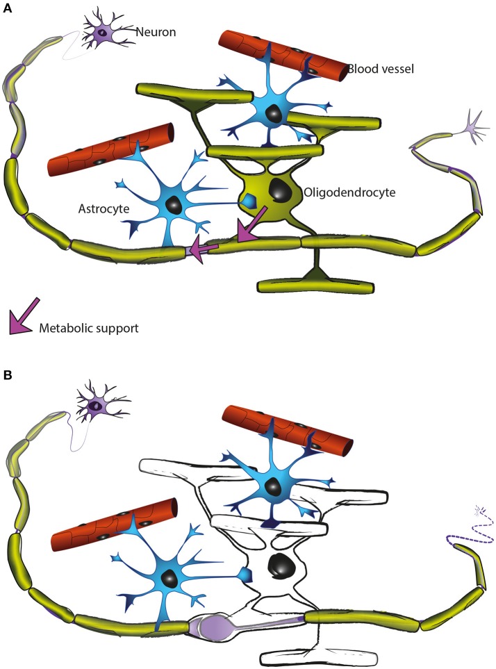
Axons are electrically excitable, cable-like neuronal processes that relay information between neurons within the nervous system and between neurons and peripheral target tissues. In the central and peripheral nervous systems, most axons over a critical diameter are enwrapped by myelin, which reduces internodal membrane capacitance and facilitates rapid conduction of electrical impulses. The spirally wrapped myelin sheath, which is an evolutionary specialisation of vertebrates, is produced by oligodendrocytes and Schwann cells; in most mammals myelination occurs during postnatal development and after axons have established connection with their targets. Myelin covers the vast majority of the axonal surface, influencing the axon's physical shape, the localisation of molecules on its membrane and the composition of the extracellular fluid (in the periaxonal space) that immerses it. Moreover, myelinating cells play a fundamental role in axonal support, at least in part by providing metabolic substrates to the underlying axon to fuel its energy requirements. The unique architecture of the myelinated axon, which is crucial to its function as a conduit over long distances, renders it particularly susceptible to injury and confers specific survival and maintenance requirements. In this review we will describe the normal morphology, ultrastructure and function of myelinated axons, and discuss how these change following disease, injury or experimental perturbation, with a particular focus on the role the myelinating cell plays in shaping and supporting the axon.

Keywords: Schwann cell; axonal transport; cytoskeleton; energy; morphology; neuroinflammation; oligodendrocyte.


Figures

Figure 1
Figure 1
Ultrastructure of myelinated axons in the CNS and PNS. (A, upper) In the CNS, myelinated axons are densely packed within white matter (here: mouse optic nerve) and the myelin sheaths of neighboring fibers often directly touch. At high magnification axonal cytoskeletal elements are visible: microtubules (arrows) and neurofilaments (arrowheads). As indicated in the schematic, mitochondria are closely associated with microtubules (M, mitochondria; IT, inner tongue; OT, outer tongue). (A, lower) In the PNS, the Schwann cell plasma membrane is covered with a basal lamina and the myelinated fibers are separated by connective tissue. Small caliber axons are not myelinated, but organized in so-called Remak bundles (R) formed by non-myelinating Schwann cells. At high magnification, mitochondria and cytoskeletal elements can be observed. (B) Schematic representation of a myelinated CNS fiber: The plasma membrane of the myelinating glial cell is depicted in green. The major dense line (depicted in grey) results from the apposition of the cytoplasmic surfaces of the plasma membrane. (C) Electron micrograph of a PNS node of Ranvier and (D, upper) schematic representation of the elements of a node: paranodal loops of the myelin sheath (green), Nav1.6 channels (red), Kv1 channels (blue), neurofilaments (arrowheads), microtubules (open arrowheads), mitochondria (M), which are transported along microtubules. N, node; PN, paranode; JPN, juxta paranode; IN, internode. (D, lower) Schematic representation of non-myelinated axon with uniform distribution of Nav1.6 channels along the axolemma.
Figure 2
Figure 2
Axonal organelle accumulations after traumatic injury or interruption of axonal transport. (A) Early description of axonal pathology after traumatic spinal cord injury in the rat, based on electron microscopic observations. (1) After transection the axonal stump swells and accumulates mitochondria, vesicles and dense bodies. (2) In a degenerative stage the organelles disintegrate. (3) A regenerative axon is characterized by a growth cone with multiple organelles. (4) A dystrophic axonal enlargement, caused by demyelinating disease or vitamin E deficiency is characterized by abundant filaments and organelles such as mitochondria, dense bodies, layered membrane loops, and cytoplasmic dense material. Reproduced from Lampert (1967), with permission. (B) Electron micrographs of focal swellings and organelle accumulations in CNS axons (upper: cross section, lower: longitudinal section), here derived from white matter of adult Plp1 null mice. Note that organelle accumulations are predominantly located in the juxtaparanodal region (lower). (C) Electron micrographs of axonal changes in the PNS after interruption of axonal transport in the mouse saphenous nerve by cooling. At the distal side of the transport block, retrogradely transported axonal elements accumulate along the internode. These are, among others, multivesicular bodies (Mv), lamellated bodies (Lb) which intermingle with microtubules (T), and neurofilaments (Upper: cross section, Lower: longitudinal section). Reproduced from Tsukita and Ishikawa (1980) with permission.
Figure 3
Figure 3
(A) Electron micrograph showing the multilayered myelin sheath of a CNS axon in cross section. Note the regular periodicity of compact myelin, which is composed of the major dense line, reflecting the fused intracellular glial surfaces (white dots), and the 2 nm wide intraperiod space between the extracellular surfaces of adjacent wraps (blue dots). Arrows point to radial components. (B) In mature CNS, a single cytoplasmic channel is present around the perimeter of the glial cell process (light green), which is best understood, if the myelin is virtually unwrapped (schematically demonstrated for one internodal segment). The cytoplasmic channel connects the glial cell soma with the most distal part of the myelin sheath and contains organelles and cytoskeletal elements, as shown in C. (C) Electron micrograph of CNS paranodal loops, abutting the axon. These contain microtubules (arrows) and vesicles (arrowheads). (A,C were reproduced from Edgar and Griffiths, , in Diffusion MRI, From Quantitative Measurement to in vivo Neuroanatomy; published by Elsevier).
Figure 4
Figure 4
Morphological characteristics of white matter in mouse “myelin mutants”. (A) Normal myelinated optic nerve axons. (B) In the shiverer mouse, oligodendrocytes lacking myelin basic protein (MBP) are not able to form compact myelin, though a few loose myelin-like wraps are observed. Note the increased oligodendrocytic processes (asterisk). AS: Astrocyte process. (C,D) In contrast, the CNPnull mouse produces compact myelin but develops pronounced axonal swellings (asterisks in C) and enlarged inner tongues (arrowheads in D).
Figure 5
Figure 5
Oligodendrocytes support axons. (A) In the CNS, oligodendrocytes (green) provide axons with the insulating myelin sheath (green: myelinated internodes). Moreover, oligodendrocytes support axonal integrity and function independent of myelination per se (for details see main text). Astrocytes (blue) not only contact blood vessels but additionally interact with axons and oligodendrocytes and contribute to brain homeostasis. Oligodendrocyte precursor cells (not illustrated) are also present in the mature CNS and are the main cellular source for new oligodendrocytes after injury and in remyelination. (B) Oligodendrocyte dysfunction leads to a perturbed axon-glia interaction which ultimately impairs axonal health. As the myelinic channel system is likely acting as a route by which oligodendrocytes supply metabolites to the myelinated axons, any perturbation of this system could potentially impact axonal integrity, resulting for example in focal axonal swelling and distal axonal degeneration.

References

    1. Adlkofer K., Martini R., Aguzzi A., Zielasek J., Toyka K. V., Suter U. (1995). Hypermyelination and demyelinating peripheral neuropathy in Pmp22-deficient mice. Nat. Genet. 11, 274–280. 10.1038/ng1195-274 - DOI - PubMed
    1. Almeida R. G., Lyons D. A. (2017). On myelinated axon plasticity and neuronal circuit formation and function. J. Neurosci. 37, 10023–10034. 10.1523/JNEUROSCI.3185-16.2017 - DOI - PMC - PubMed
    1. Andrews H. E., White K., Thomson C. E., Edgar J. M., Bates D., Griffiths I. R., et al. . (2006). Increased axonal mitochondrial activity as an adaptation to myelin deficiency in the shiverer mouse. J. Neurosci. Res. 83, 1533–1539. 10.1002/jnr.20842 - DOI - PubMed
    1. Arancibia-Carcamo I. L., Ford M. C., Cossell L., Ishida K., Tohyama K., Attwell D. (2017). Node of Ranvier length as a potential regulator of myelinated axon conduction speed. Elife 6:e23329. 10.7554/eLife.23329 - DOI - PMC - PubMed
    1. Arthur-Farraj P. J., Latouche M., Wilton D. K., Quintes S., Chabrol E., Banerjee A., et al. . (2012). c-Jun reprograms Schwann cells of injured nerves to generate a repair cell essential for regeneration. Neuron 75, 633–647. 10.1016/j.neuron.2012.06.021 - DOI - PMC - PubMed

LinkOut - more resources