. 2018 Jul 12:11:235.
doi: 10.3389/fnmol.2018.00235. eCollection 2018.

Beneficial Effect of a Selective Adenosine A2A Receptor Antagonist in the APPswe/PS1dE9 Mouse Model of Alzheimer's Disease

Affiliations

Beneficial Effect of a Selective Adenosine A2A Receptor Antagonist in the APPswe/PS1dE9 Mouse Model of Alzheimer's Disease

Emilie Faivre et al. Front Mol Neurosci. .

Abstract

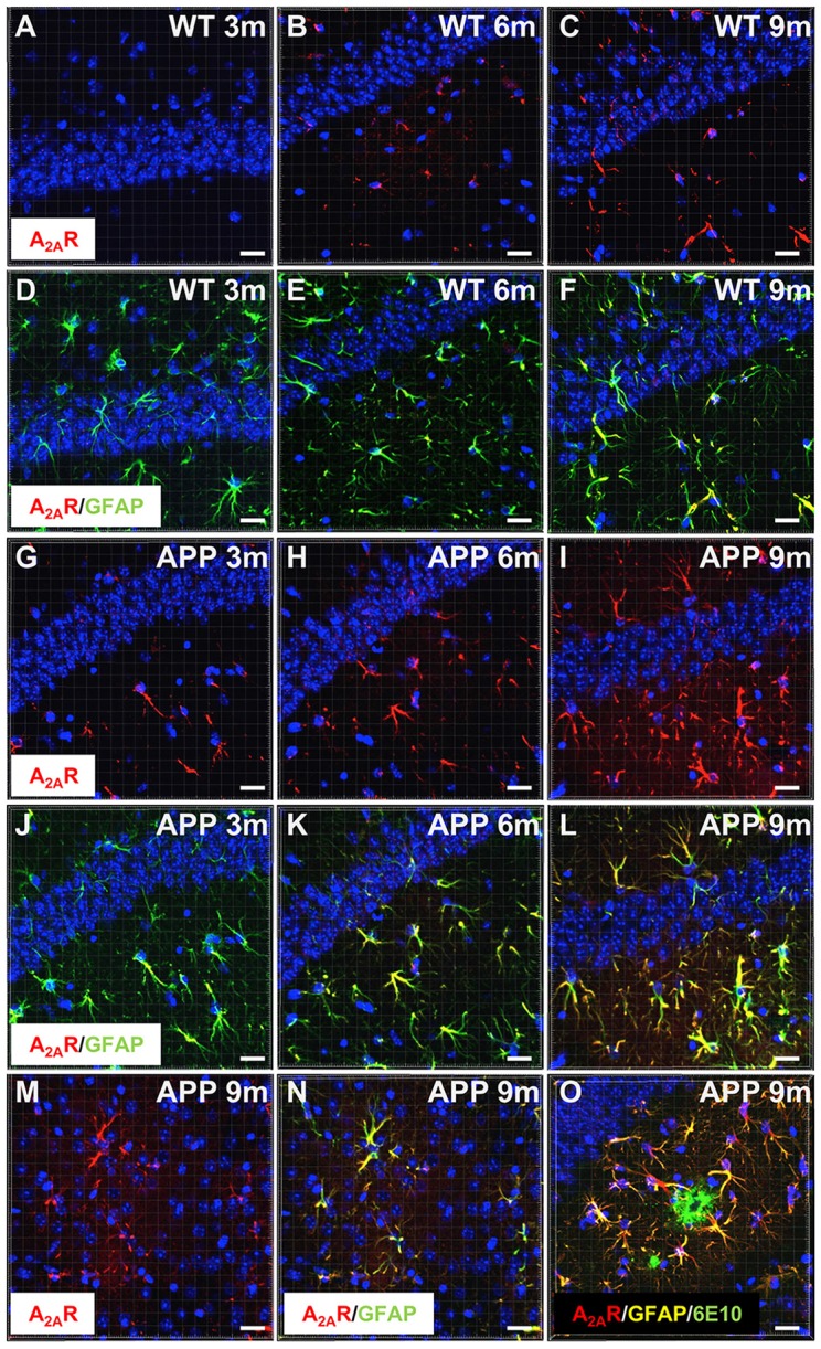
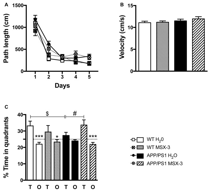
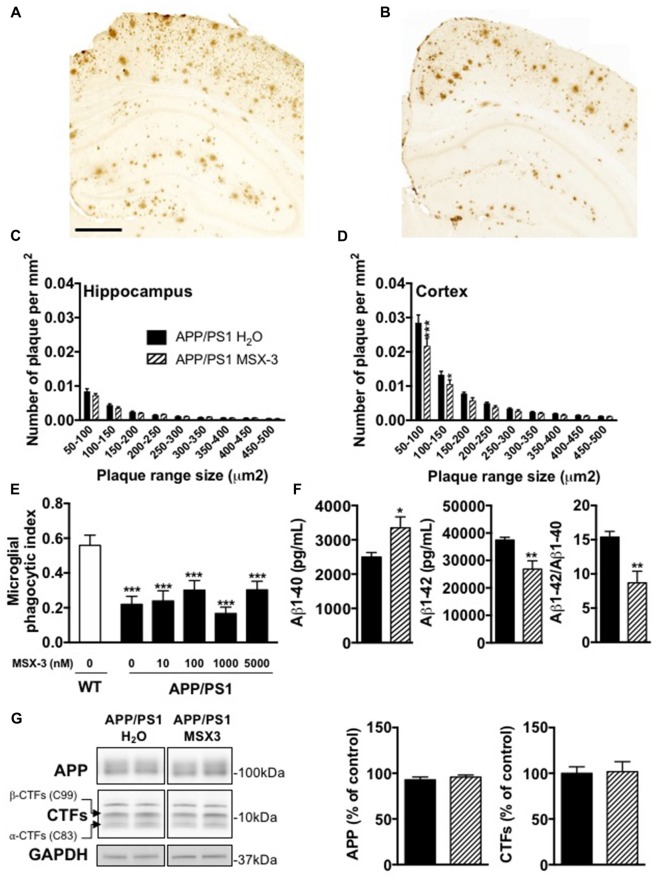
Consumption of caffeine, a non-selective adenosine A2A receptor (A2AR) antagonist, reduces the risk of developing Alzheimer's disease (AD) and mitigates both amyloid and Tau lesions in transgenic mouse models of the disease. While short-term treatment with A2AR antagonists have been shown to alleviate cognitive deficits in mouse models of amyloidogenesis, impact of a chronic and long-term treatment on the development of amyloid burden, associated neuroinflammation and memory deficits has never been assessed. In the present study, we have evaluated the effect of a 6-month treatment of APPsw/PS1dE9 mice with the potent and selective A2AR antagonist MSX-3 from 3 to 9-10 months of age. At completion of the treatment, we found that the MSX-3 treatment prevented the development of memory deficits in APP/PS1dE9 mice, without significantly altering hippocampal and cortical gene expressions. Interestingly, MSX-3 treatment led to a significant decrease of Aβ1-42 levels in the cortex of APP/PS1dE9 animals, while Aβ1-40 increased, thereby strongly affecting the Aβ1-42/Aβ1-40 ratio. Together, these data support the idea that A2AR blockade is of therapeutic value for AD.

Keywords: A2A; Alzheimer’s disease; adenosine receptor; amyloid; memory.


Figures

Figure 1
Figure 1
Astrocytic upsurge of A2ARs in APP/PS1 mice. Representative photomicrographs of hippocampal immunostaining for the A2A receptor (A2AR; red) (A–C,G–I) and merged with the astrocyte marker GFAP (green) (D–F,J–L) from WT mice (A–F) and the APP/PS1 mice (G–L), at different ages: 3 months (A,D,G,J), 6 months (B,E,H,K), and 9–10 months (C,F,I,L). Representative photomicrographs of A2AR expression (red) (M) and the merged with GFAP marker (green; N) in the cortex of 9-month-old APP/PS1 mice. Representative photomicrograph of A2AR expression (red) GFAP (yellow) and 6E10-positive amyloid plaque marker (green) in the hippocampus of 9 month-old APP/PS1 mice (O). Cell nuclei were labeled with DAPI (blue). Scale bar = 20 μm.
Figure 2
Figure 2
A2AR blockade prevents spatial memory impairments in APP/PS1 transgenic mice. Effect of MSX-3 treatment on spatial learning and memory using the Morris water-maze task. (A) Learning, as indicated by the equivalent path length needed to find the hidden platform (PF). At day 2, APP/PS1 mice exhibited a slight but significant higher path length as compared to WT animals (***p < 0.001 vs. WT H2O using One-way analysis of variance (ANOVA) followed by LSD Fisher post hoc test). (B) All genotypes exhibited a comparable velocity in the maze, suggesting no motor deficits. (C) Spatial memory was assessed 72 h after the last day of learning. Results represent the percentage of time spent in the target (T) vs. non-target (O) quadrants. WT mice (both treated with water or MSX-3) spent significantly more time in the T quadrant, indicative of a preserved spatial memory. While APP/PS1 mice exhibited spatial memory deficits as underlined by their lack of preference for the T quadrant, APP/PS1-MSX-3 treated mice behaved as WT mice, suggesting a rescue of memory impairment. *p < 0.05 ***p < 0.001 T vs. O; $p < 0.05 WT vs. APP/PS1; #p < 0.05 APP/PS1-H2O vs. APP/PS1 MSX-3 using One-way ANOVA followed by LSD Fisher post hoc test; N = 12–16 per group; Results are expressed as mean ± SEM.
Figure 3
Figure 3
Impact of MSX-3 treatment on amyloid load, Aβ levels and phagocytic capacity of microglia in APP/PS1 mice. Representative images of 6E10 staining in the brains of 10 months old APP/PS1 treated with water (A) or MSX-3 (B). Scale bar = 500 μm. Distribution of amyloid plaques size were examined in the hippocampus (C) and cortex (D) of APP/PS1 mice. We found that the treatment with MSX-3 significantly reduced, in the cortex (D) but not in the hippocampus (C), the density of plaques of lower size (between 50–150 μm2) as compared with APP/PS1 H2O animals (50–150 μm2, p < 0.001; 100–150 μm2, p < 0.05 vs. APP/PS1 H2O using Two-Way ANOVA followed by LSD Fisher post hoc test; N = 7–9/group). (E) Effect of acute MSX-3 treatment on microglial phagocytic index determined in an in situ live cerebral slice assay of 12-month-old APP/PS1-Csf1r-EGFP mice and Csf1r-EGFP wildtype littermates after 60 min pre-incubation with the indicated concentration of MSX-3. Datasets were tested for significance with the One-way ANOVA and represent data from three independent experiments. ***p < 0.001 vs. WT (F) As measured by ELISA, MSX-3 treatment decreased Aβ1–42 levels in the cortex of APP/PS1 mice while Aβ1–40 levels was found increased. Overall, the Aβ1–42/Aβ1–40 ratio was found significantly reduced by the A2A antagonist treatment (*p < 0.05, **p < 0.01 vs. APP/PS1 H2O using Student’s t-test; N = 7–11/group). (G) Western blot analysis performed in cortex of water and MSX-3 treated APP/PS1dE9 mice did not revealed any change in APP and Carboxyterminal fragments (CTFs) expression (N = 6/group). Results are expressed as mean ± SEM.

References

    1. Abbott L. F., Regehr W. G. (2004). Synaptic computation. Nature 431, 796–803. 10.1038/nature03010 - DOI - PubMed
    1. Arendash G. W., Mori T., Cao C., Mamcarz M., Runfeldt M., Dickson A., et al. . (2009). Caffeine reverses cognitive impairment and decreases brain amyloid-β levels in aged Alzheimer’s disease mice. J. Alzheimers Dis. 17, 661–680. 10.3233/JAD-2009-1087 - DOI - PubMed
    1. Arendash G. W., Schleif W., Rezai-Zadeh K., Jackson E. K., Zacharia L. C., Cracchiolo J. R., et al. . (2006). Caffeine protects Alzheimer’s mice against cognitive impairment and reduces brain-amyloid production. Neuroscience 142, 941–952. 10.1016/j.neuroscience.2006.07.021 - DOI - PubMed
    1. Baeta-Corral R., Johansson B., Giménez-Llort L. (2018). Long-term treatment with low-dose caffeine worsens BPSD-like profile in 3xTg-AD mice model of Alzheimer’s disease and affects mice with normal aging. Front. Pharmacol. 9:79. 10.3389/fphar.2018.00079 - DOI - PMC - PubMed
    1. Batalha V. L., Ferreira D. G., Coelho J. E., Valadas J. S., Gomes R., Temido-Ferreira M., et al. . (2016). The caffeine-binding adenosine A2A receptor induces age-like HPA-axis dysfunction by targeting glucocorticoid receptor function. Sci. Rep. 6:31493. 10.1038/srep31493 - DOI - PMC - PubMed