. 2018 Jul 11:9:1544.
doi: 10.3389/fmicb.2018.01544. eCollection 2018.

The Immune Protection Induced by a Serine Protease Inhibitor From the Foodborne Parasite Trichinella spiralis

Affiliations

The Immune Protection Induced by a Serine Protease Inhibitor From the Foodborne Parasite Trichinella spiralis

Yan Y Song et al. Front Microbiol. .

Abstract

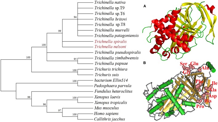
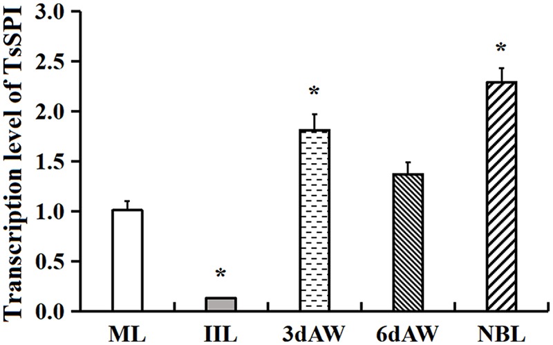
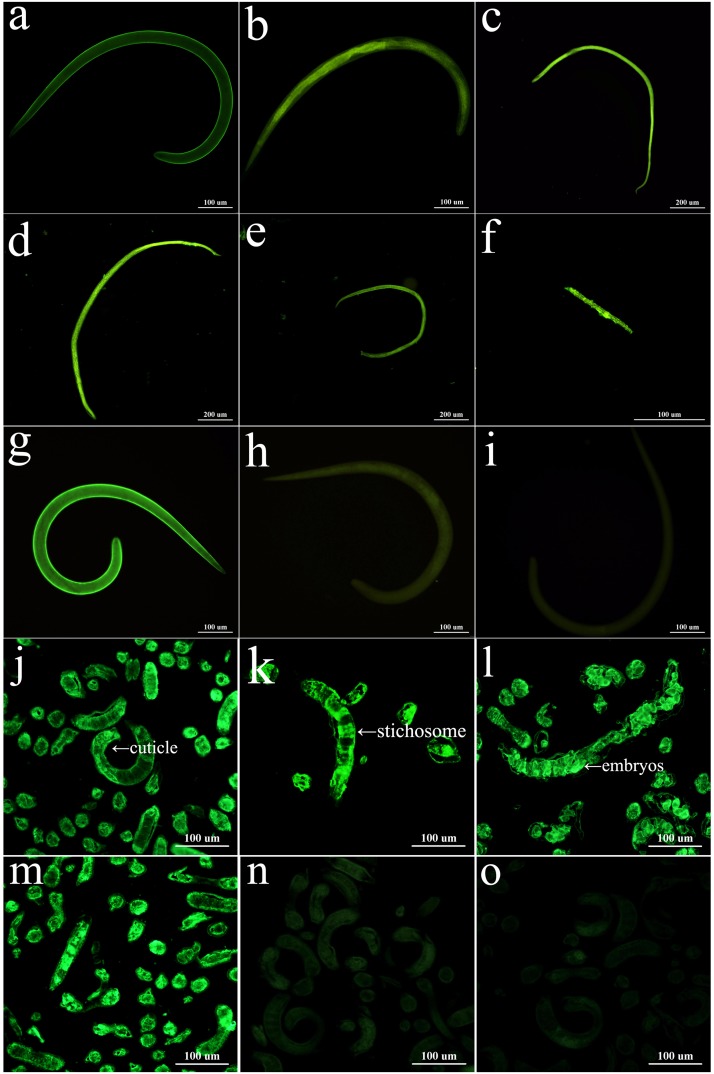
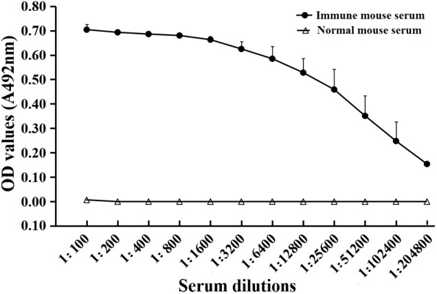
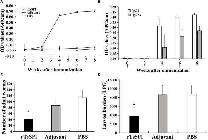
Serine protease inhibitors (SPI) are a superfamily of the proteins able to suppress serine protease activity, and may exert the major biological function in complement activation, inflammation, and fibrinolysis. A SPI was identified from Trichinella spiralis adult worms (AW) by immunoproteomics with early infection sera. The aim of this study was to investigate the protective immune elicited by TsSPI. The complete TsSPI cDNA sequence was cloned into pQE-80 L and then expressed in Escherichia coli BL21. The rTsSPI was purified and its antigenicity was determined by Western blotting analysis. By using anti-rTsSPI serum the native TsSPI was identified in somatic and ES proteins from muscle larvae (ML). The results of qPCR and immunofluorescence assay (IFA) revealed that the expression of the TsSPI gene was observed throughout all developmental stages of T. spiralis (ML, intestinal infective larvale, 3- and 6-days AW, and newborn larvae, NBL), located principally in cuticles, stichosome, and embryos of this parasitic nematode. Vaccination of mice with rTsSPI triggered high level of anti-TsSPI IgG response, and showed a 62.2 and 57.25% worm burden reduction in the recovery of intestinal AW at 6 days post-infection (dpi) and ML at 35 dpi, respectively. The TsSPI might be a novel potential target for anti-Trichinella vaccine.

Keywords: Trichinella spiralis; identification; immune protection; serine protease inhibitors; tissue localization.


Figures

FIGURE 1
FIGURE 1
Phylogenetic trees of serine protease inhibitors (SPI) of 22 organisms with the NJ method and mapped with MEGA (left) and the predicted three-dimensional structure of T. spiralis TsSPI protein (right). (A) The predicted three-dimensional structure of TsSPI protein contains 9 α-helixes (in red) and 12 β-strand (in yellow). (B) The functional domain with an active site carrying a classic SPI reactive central loop (RCL) consisted of ten amino acids. The active site of TsSPI is marked on pink.
FIGURE 2
FIGURE 2
Identification of rTsSPI. (A) SDS–PAGE analysis of the rTsSPI. Lane M: molecular weight markers; Lane 1: recombinant bacteria lysate before induction; Lane 2: recombinant bacteria lysate after IPTG induction; Lane 3: the purified rTSPI. (B) SDS–PAGE of somatic proteins (Lane 1), ES proteins (Lane 2), and rTspGST (Lane 3). (C) Western blotting. The native TsSPI protein in ML somatic proteins (lane 1) and ES proteins (lane 2) as well as the rTsSPI (lane 3) were identified by anti-rTsSPI serum. The ML somatic proteins (lane 4), ES proteins (lane 5) and rTsSPI (lane 6) were also probed with serum samples of T. spiralis-infected mice. The ML somatic proteins (lane 7), ES proteins (lane 8), and rTsSPI (lane 9) were not recognized with pre-immune sera of normal mice.
FIGURE 3
FIGURE 3
qPCR analysis of TsSPI transcription level at various stages of T. spiralis. The TsSPI mRNAs from ML, IIL, 3- and 6-days AW, and NBL were extracted and amplified by qPCR. The TsSPI transcription level was calculated according to the Ct (2-ΔΔCt) method. The fold change in the TsSPI genes was normalized to G3PDH used as a housekeeping gene control. The data shown are representative from three independent experiment. Statistical differences are marked with asterisks () compared with the ML stage at P < 0.05.
FIGURE 4
FIGURE 4
Expression and immunolocalization of TsSPI in different T. spiralis phases. (a–i) The intact worms were examined by IFA with different mouse sera. When the anti-rTsSPI serum was used, immunostaining was observed on the surface of ML (a), IIL (b), 3-day female adult (c), 6-day female (d), 6-day male adult (e), and NBL (f). ML recognized by infection serum (g) was used as a positive serum control; ML incubated using pre-immune serum (h) and PBS (i) were used as negative serum and blank controls. (j–o): Worm sections were examined by IFA with different mouse sera. The worm sections were probed by anti-rTsSPI serum, intense fluorescent staining is observed in cuticles, stichosome of ML (j), IIL (k), and embryos of 3-day female adult (i). The ML reacted by infection serum (m) was used as a positive serum control; there are no staining in the ML with pre-immune serum (n) and PBS (o) as a negative control. Scale-bars: 100 μm.
FIGURE 5
FIGURE 5
Serum anti-rTsSPI IgG titers after the fourth immunization in immunized mice determined by ELISA. The data are presented as the mean OD values ± SD of the antibody levels from twenty mice.
FIGURE 6
FIGURE 6
Protective immunity in mice immunized with rTsSPI. Serum IgG (A) and IgG subtype (B) responses to the rTsSPI at different times post immunization were measured by ELISA. The data are presented as the mean OD values ± SD of specific anti-rTsSPI antibodies from ten mice. The arrow (↑) represents immunization times. The number of adults recovered from intestines (C) and larvae per gram (LPG) of muscles (D) in immunized mice after challenge with 300 T. spiralis muscle larvae. Results are expressed as the mean ± SD of 10 mice per group. An obvious reduction was observed in the worm burdens of intestinal adults at 6 dpi and muscle larvae at 35 dpi (P < 0.01).

References

    1. Apweiler R., Bairoch A., Wu C. H., Barker W. C., Boeckmann B., Ferro S., et al. (2004). UniProt: the universal protein knowledgebase. Nucleic Acids Res. 32 D115–D119. 10.1093/nar/gkh131 - DOI - PMC - PubMed
    1. Bai X., Hu X. X., Liu X. L., Tang B., Liu M. Y. (2017). Current research of trichinellosis in China. Front. Microbiol. 8:1472 10.3389/fmicb.2017.01472 - DOI - PMC - PubMed
    1. Bien J., Cabaj W., Moskwa B. (2015). Proteomic analysis of potential immunoreactive proteins from muscle larvae and adult worms of Trichinella spiralis in experimentally infected pigs. Folia Parasitol. 62:022. 10.14411/fp.2015.022 - DOI - PubMed
    1. Bioreau P., Vayssier M., Fabien J. F., Perret C., Calamel M., Soule C. (1997). Characterization of eleven antigenic groups in Trichinella genus and identification of stage and species markers. Parasitology 115 641–651. 10.1017/S0031182097001716 - DOI - PubMed
    1. Blanton R. E., Licate L. S., Aman R. A. (1994). Characterization of a native and recombinant Schistosoma haematobium serine protease inhibitor gene product. Mol. Biochem. Parasitol. 63 1–11. 10.1016/0166-6851(94)90003-5 - DOI - PubMed

LinkOut - more resources