Review
. 2018 Jun;7(3):368-375.
doi: 10.21037/tlcr.2018.06.05.

Extrapulmonary neoplasms in lung cancer screening

Affiliations
Review

Extrapulmonary neoplasms in lung cancer screening

Myrna C B Godoy et al. Transl Lung Cancer Res. 2018 Jun.

Abstract

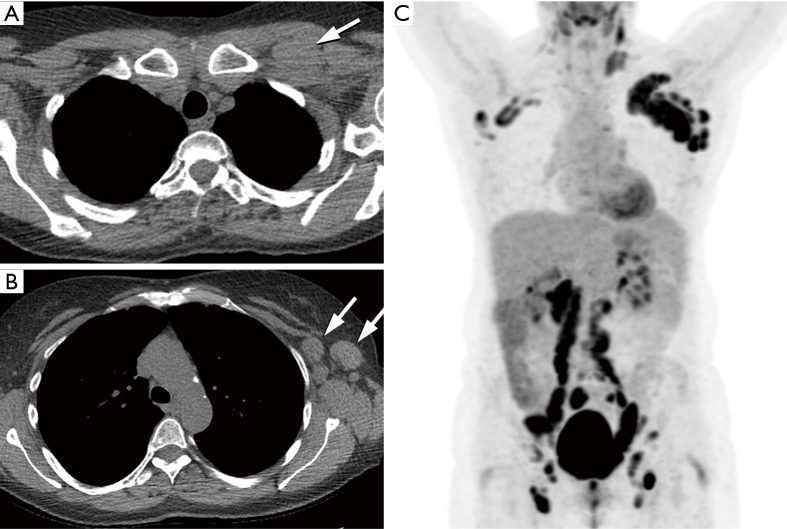
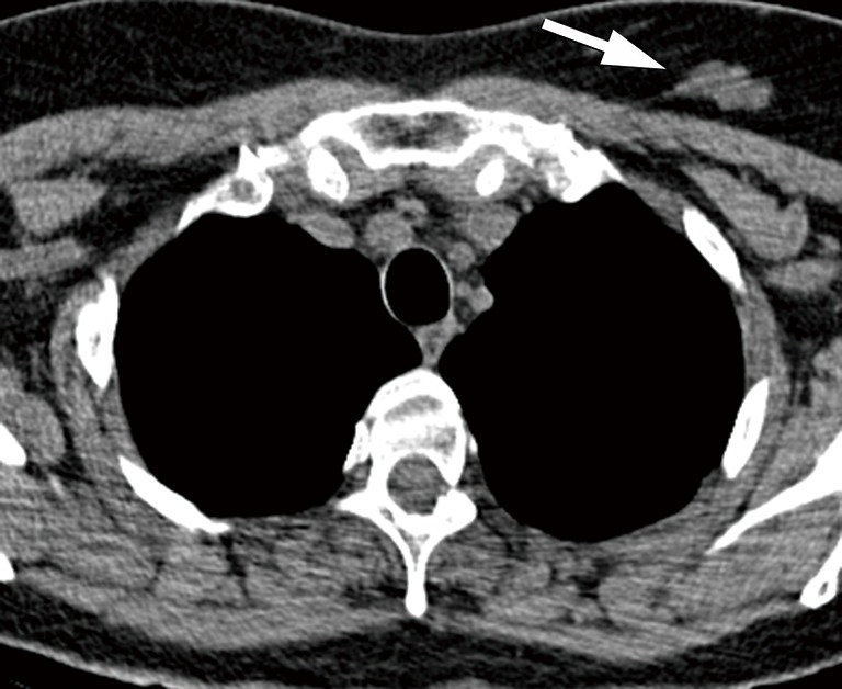
A significant reduction in lung cancer specific mortality rate was demonstrated by the National Lung Screening Trial (NLST) in participants who had annual low-dose computed tomography (LDCT) screening. In addition to early detection of lung cancer, lung cancer screening (LCS) provides an opportunity to detect cardiovascular disease, pulmonary disease (such as chronic obstructive pulmonary disease and interstitial lung disease), and extrapulmonary neoplasms, such as thyroid, breast, kidney, liver, esophageal, pancreatic and mediastinal tumors. Considering the fact that 22.3% of the certified deaths in the computed tomography (CT) arm of the NLST trial were due to extrapulmonary malignancies, compared to 22.9% of deaths from lung cancer, it is possible that early diagnosis and treatment of clinically significant incidental findings may further decrease morbidity and mortality in screening participants. In this article we review prevalence, clinical relevance and management of incidentally detected extrapulmonary malignancies.

Keywords: Lung cancer; computed tomography (CT); incidental findings (IFs); low-dose computed tomography (LDCT); neoplasm; screening.


Conflict of interest statement

Conflicts of Interest: The authors have no conflicts of interest to declare

Figures

Figure 1
Figure 1
Mediastinal incidentaloma. LDCT shows a 2-cm mediastinal soft-tissue nodule suspicious for thymic neoplasm (arrow). The lesion was stable at 3-month follow-up LDCT (not shown) and subsequently managed with annual LDCT follow-up. LDCT, low-dose computed tomography.
Figure 2
Figure 2
Esophageal tumor. Screening LDCT at baseline (A) and year 1 (B) shows focal circumferential wall thickening in the distal esophagus (arrows) which has increased. Biopsy confirmed esophageal adenocarcinoma. LDCT, low-dose computed tomography.
Figure 3
Figure 3
Lymphoma. Baseline LDCT images (A,B) show left retropectoral and axillary lymphadenopathy (arrows) measuring up to 3.1 cm × 2.2 cm . Further investigation was performed with ultrasound and fine needle aspiration. Pathology confirmed follicular lymphoma; (C) MIP coronal PET/CT image performed for staging shows hypermetabolic multicompartmental lymphadenopathy involving the left neck and bilateral axillary, retroperitoneal, iliac, inguinal and femoral regions. MIP, intensity projection image; LDCT, low-dose computed tomography.
Figure 4
Figure 4
Breast incidentaloma. LDCT shows a soft-tissue mass in the left breast (arrow). Management recommendation includes correlation with mammography and/or ultrasonography. Breast cancer was confirmed on pathology. LDCT, low-dose computed tomography.
Figure 5
Figure 5
Adrenal gland nodule. LDCT shows a 1.3-cm right adrenal gland nodule (arrow). Patient had no prior imaging and no history of malignancy. For incidental adrenal lesions measuring 1–2 cm with no prior imaging in patients with no cancer history, evaluation for lesion stability at the 12-month LDCT follow-up may be sufficient. Follow-up at subsequent annual LDCT screening showed no significant interval change (not shown), most consistent with an adenoma. LDCT, low-dose computed tomography.
Figure 6
Figure 6
Solid pancreatic mass. LDCT shows a poorly-marginated mass in the head of the pancreas (asterisk), consistent with pancreatic cancer. Pancreatic adenocarcinoma was confirmed on pathology. LDCT, low-dose computed tomography.
Figure 7
Figure 7
Cystic pancreatic lesion. (A) LDCT shows an indeterminate 2-cm low-attenuation lesion identified in the tail of the pancreas (arrow); (B) pancreas protocol CT imaging performed with intravenous contrast allows better characterization of the well-defined cystic lesion in the tail of the pancreas (arrow). Endoscopic ultrasound/fine needle aspiration (EUS/FNA) showed no malignant cells. The lesion remained stable on subsequent follow-up imaging (not shown). LDCT, low-dose computed tomography.

References

    1. National Lung Screening Trial Research Team , Aberle DR, Adams AM, et al. Reduced lung-cancer mortality with low-dose computed tomographic screening. N Engl J Med 2011;365:395-409. 10.1056/NEJMoa1102873 - DOI - PMC - PubMed
    1. Nguyen XV, Davies L, Eastwood JD, et al. Extrapulmonary Findings and Malignancies in Participants Screened With Chest CT in the National Lung Screening Trial. J Am Coll Radiol 2017;14:324-30. 10.1016/j.jacr.2016.09.044 - DOI - PubMed
    1. Kucharczyk MJ, Menezes RJ, McGregor A, et al. Assessing the impact of incidental findings in a lung cancer screening study by using low-dose computed tomography. Can Assoc Radiol J 2011;62:141-5. 10.1016/j.carj.2010.02.008 - DOI - PubMed
    1. MacRedmond R, Logan PM, Lee M, et al. Screening for lung cancer using low dose CT scanning. Thorax 2004;59:237-41. 10.1136/thx.2003.008821 - DOI - PMC - PubMed
    1. Swensen SJ, Jett JR, Sloan JA, et al. Screening for lung cancer with low-dose spiral computed tomography. Am J Respir Crit Care Med 2002;165:508-13. 10.1164/ajrccm.165.4.2107006 - DOI - PubMed

LinkOut - more resources