Review
. 2015;2(1):3.
doi: 10.1186/s41205-016-0004-x. Epub 2016 Sep 13.

3D printing in medicine of congenital heart diseases

Affiliations
Review

3D printing in medicine of congenital heart diseases

Shi-Joon Yoo et al. 3D Print Med. 2015.

Abstract

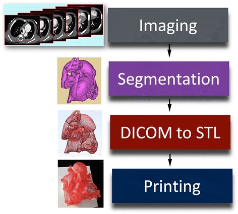
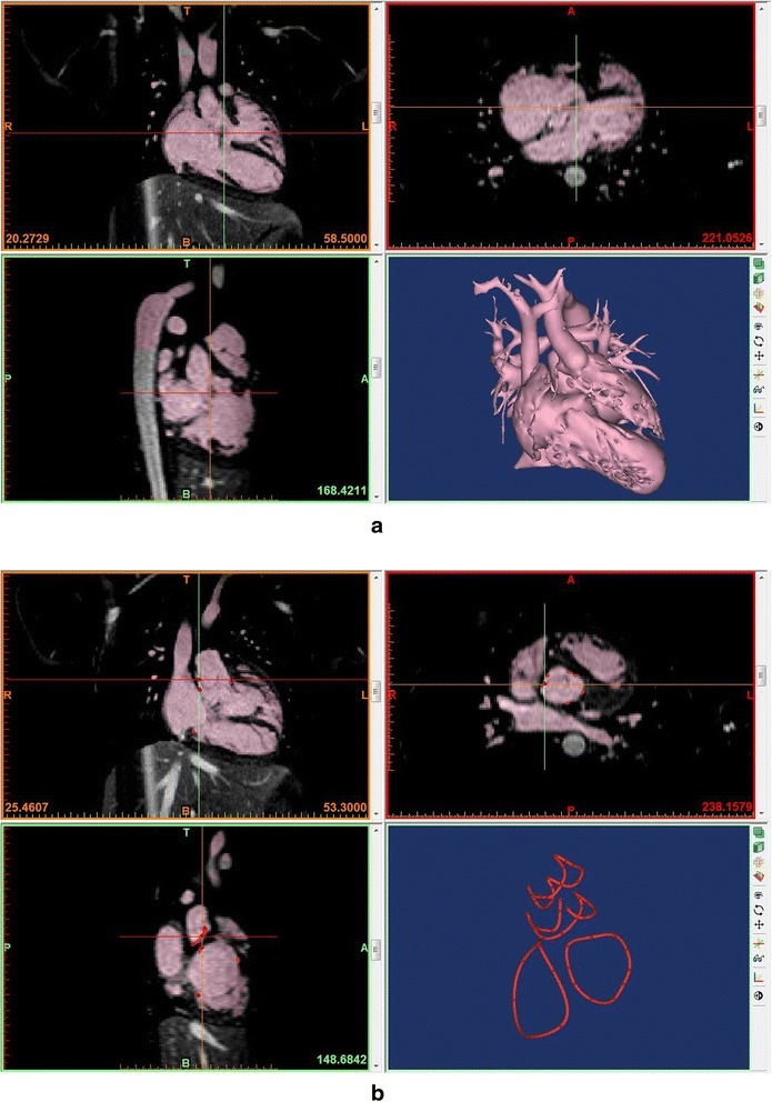
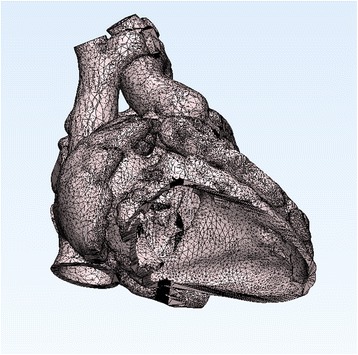
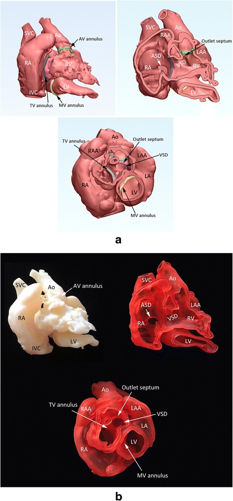
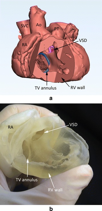
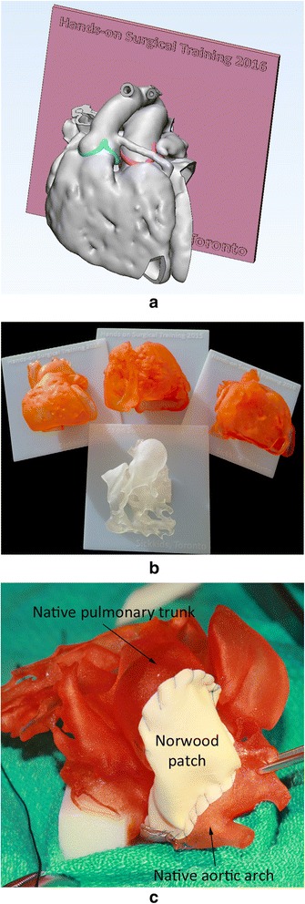
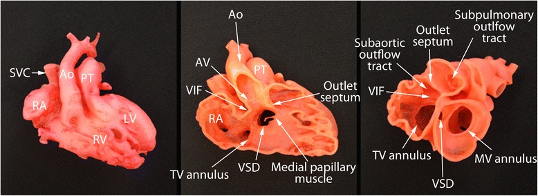
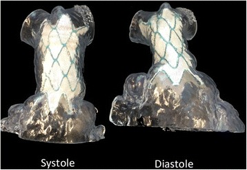
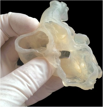
Congenital heart diseases causing significant hemodynamic and functional consequences require surgical repair. Understanding of the precise surgical anatomy is often challenging and can be inadequate or wrong. Modern high resolution imaging techniques and 3D printing technology allow 3D printing of the replicas of the patient's heart for precise understanding of the complex anatomy, hands-on simulation of surgical and interventional procedures, and morphology teaching of the medical professionals and patients. CT or MR images obtained with ECG-gating and breath-holding or respiration navigation are best suited for 3D printing. 3D echocardiograms are not ideal but can be used for printing limited areas of interest such as cardiac valves and ventricular septum. Although the print materials still require optimization for representation of cardiovascular tissues and valves, the surgeons find the models suitable for practicing closure of the septal defects, application of the baffles within the ventricles, reconstructing the aortic arch, and arterial switch procedure. Hands-on surgical training (HOST) on models may soon become a mandatory component of congenital heart disease surgery program. 3D printing will expand its utilization with further improvement of the use of echocardiographic data and image fusion algorithm across multiple imaging modalities and development of new printing materials. Bioprinting of implants such as stents, patches and artificial valves and tissue engineering of a part of or whole heart using the patient's own cells will open the door to a new era of personalized medicine.

Keywords: 3D printing; Congenital heart disease; Surgical simulation; Surgical training.


Figures

Fig. 1
Fig. 1
Diagram showing the steps in data processing for 3D printing. DICOM, digital imaging and communication in medicine; STL, stereolithography, standard tessellation language or standard triangle language
Fig. 2
Fig. 2
Segmentation process in a commercially available software program (Mimics®, Materialise, Leuven, Belgium) using magnetic resonance angiograms from a patient with congenitally corrected transposition of the great arteries with a ventricular septal defect. a Segmentation using thresholding and manual edition with a volume rendered image on the right lower panel. b Linear representation of the cardiac valvar attachments. A few points of attachment sites of each cardiac valve were marked and connected using a tool called “spline”
Fig. 3
Fig. 3
Surface geometry in STL (Stereolithography or Standard Tessellation Language) file from the patient shown in Fig. 2. The surface of the object is divided into a number of triangles without any gap and overlap
Fig. 4
Fig. 4
Screen-display of the STL files (a) and photographs of the corresponding models (b) in a case with so-called twisted or criss-cross heart with transposition of the great arteries and a ventricular septal defect (VSD). Cast model (top left), wall model after removal of the anterior free wall of the right atrium and right and left ventricles (top right), and wall model with the apical two thirds of the ventricles removed (bottom) are shown. The cardiac valve annuli are marked by the spline curves that were graphically designed as shown in Fig. 2B. Ao, aorta; ASD, atrial septal defect; AV, aortic valve; IVC, inferior vena cava; LA, left atrium; LAA, left atrial appendage; LV, left ventricle; MV, mitral valve; SVC, superior vena cava; TV, tricuspid valve
Fig. 5
Fig. 5
Screen-display (a) and photograph (b) of the 3D print model of the heart with tetralogy of Fallot. The full thickness of the myocardium was carefully segmented with both endocardial and epicardial boundaries delineated by thresholding and manual editing. Although it is considered ideal, the postprocessing was time consuming and the model is stiff to be used for surgical simulation. Ao, aorta; PT, pulmonary trunk; SVC, superior vena cava; TV, tricuspid valve; RA, right atrium; RV, right ventricle; VSD, ventricular septal defect
Fig. 6
Fig. 6
Models for surgical practice or training. a Screen-display of the STL file shows the heart mounted on a graphically designed platform. b Four example models for surgical training. c A 3D print model with hypoplastic left heart syndrome on which a surgeon underwent a Norwood procedure
Fig. 7
Fig. 7
Photographs of the 3D print models of a case with double outlet right ventricle. Although echocardiograms showed that the ventricular septal defect (VSD) is remote from both arterial valves, 3D models show that the VSD is able to be routed to the aortic valve allowing biventricular repair. Ao, aorta; AV, aortic valve; LA, left atrium; LV, left ventricle; MV, mitral valve; SVC, superior vena cava; TV, tricuspid valve; VIF, ventriculoinfundibular fold
Fig. 8
Fig. 8
Photographs of the 3D print models of right ventricular outflow tract obtained in systole and diastole from a patient with severe pulmonary regurgitation after repair of tetralogy of Fallot. The fitness of the stent in the outflow tract was tested for both systole and diastole before the procedure
Fig. 9
Fig. 9
Photograph of a model made of silicone using injection molding technique

References

    1. Dolk H, Loane M, Garne E. Congenital heart defects in Europe: prevalence and perinatal mortality, 2000 to 2005. Circulation. 2011;123:841–9. doi: 10.1161/CIRCULATIONAHA.110.958405. - DOI - PubMed
    1. Giroud JM, Jacobs JP, Spicer D, Backer C, Martin GR, Franklin RC, Béland MJ, Krogmann ON, Aiello VD, Colan SD, Everett AD, William Gaynor J, Kurosawa H, Maruszewski B, Stellin G, Tchervenkov CI, Walters HL, III, Weinberg P, Anderson RH, Elliott MJ. Report from the international society for nomenclature of paediatric and congenital heart disease: creation of a visual encyclopedia illustrating the terms and definitions of the international pediatric and congenital cardiac code. World J Pediatr Congenit Heart Surg. 2010;1(3):300–13. doi: 10.1177/2150135110379622. - DOI - PubMed
    1. Mitsouras D, Liacouras P, Imanzadeh A, Giannopoulos AA, Cai T, Kumamaru KK, George E, Wake N, Caterson EJ, Pomahac B, Ho VB, Grant GT, Rybicki FJ. Medical 3D printing for the radiologist. Radiographics. 2015;35(7):1965–88. doi: 10.1148/rg.2015140320. - DOI - PMC - PubMed
    1. Matsumoto JS, Morris JM, Foley TA, Williamson EE, Leng S, McGee KP, Kuhlmann JL, Nesberg LE, Vrtiska TJ. Three-dimensional physical modeling: applications and experience at mayo clinic. Radiographics. 2015;35(7):1989–2006. doi: 10.1148/rg.2015140260. - DOI - PubMed
    1. Binder TM, Moertl D, Mundigler G, Rehak G, Franke M, Delle-Karth G, Mohl W, Baumgartner H, Maurer G. Stereolithographic biomodeling to create tangible hard copies of cardiac structures from echocardiographic data: in vitro and in vivo validation. J Am Coll Cardiol. 2000;35:230–7. doi: 10.1016/S0735-1097(99)00498-2. - DOI - PubMed