. 2015;2(1):5.
doi: 10.1186/s41205-016-0008-6. Epub 2016 Nov 25.

Medical 3D printing for vascular interventions and surgical oncology: a primer for the 2016 radiological society of North America (RSNA) hands-on course in 3D printing

Affiliations

Medical 3D printing for vascular interventions and surgical oncology: a primer for the 2016 radiological society of North America (RSNA) hands-on course in 3D printing

Leonid Chepelev et al. 3D Print Med. 2015.

Abstract

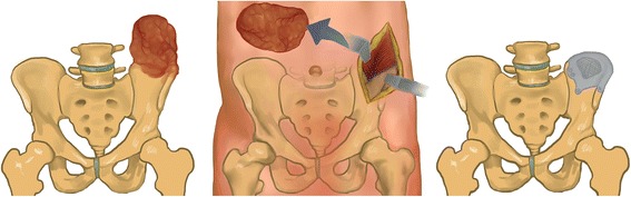
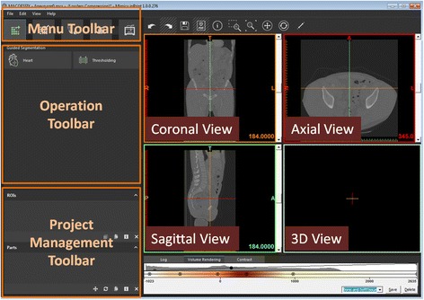
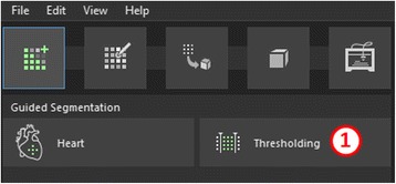
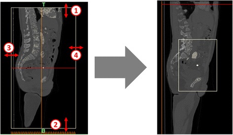
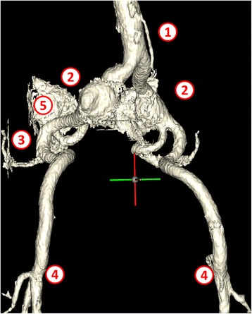
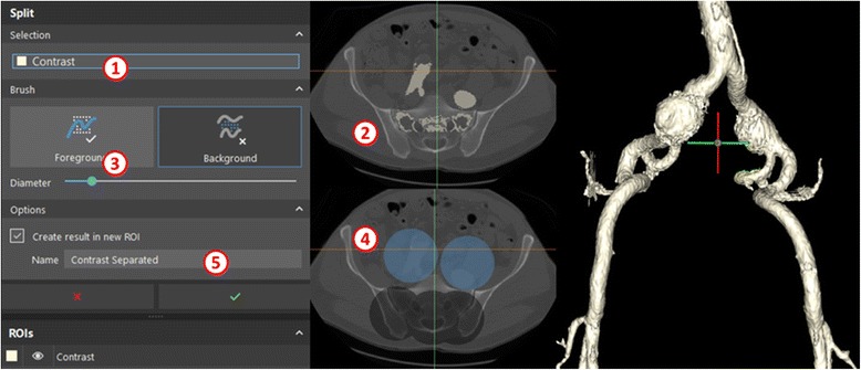
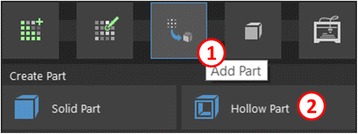
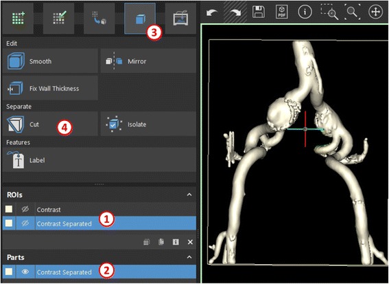
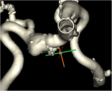
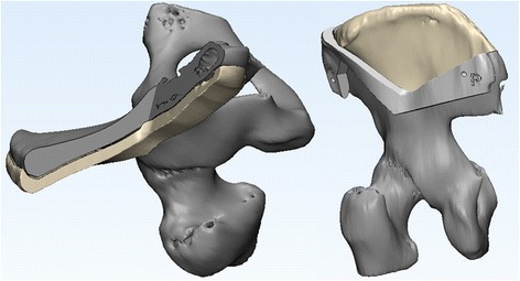
Medical 3D printing holds the potential of transforming personalized medicine by enabling the fabrication of patient-specific implants, reimagining prostheses, developing surgical guides to expedite and transform surgical interventions, and enabling a growing multitude of specialized applications. In order to realize this tremendous potential in frontline medicine, an understanding of the basic principles of 3D printing by the medical professionals is required. This primer underlines the basic approaches and tools in 3D printing, starting from patient anatomy acquired through cross-sectional imaging, in this case Computed Tomography (CT). We describe the basic principles using the relatively simple task of separation of the relevant anatomy to guide aneurysm repair. This is followed by exploration of more advanced techniques in the creation of patient-specific surgical guides and prostheses for a patient with extensive pleomorphic sarcoma using Computer Aided Design (CAD) software.

Keywords: 3D Printing; Aneurysm repair; Cancer; Computer-aided design; Implant; Orthopedic Surgery; Precision Medicine; Radiological Society of North America; Segmentation; Surgical Guide.


Conflict of interest statement

The authors declare that they have no competing interests.

Figures

Fig. 1
Fig. 1
Simplified overview of pleomorphic sarcoma supported in this work. After identifying the extent of the neoplasm (left, red), it is resected with wide margins. The skeletal defect is filled using a patient-specific implant (right)
Fig. 2
Fig. 2
Overview of the Mimics inPrint project screen
Fig. 3
Fig. 3
Thresholding function (1) within the Guided Segmentation menu
Fig. 4
Fig. 4
Settings for minimal (1) and maximal (2) attenuation thresholds, naming the ROI (3), and options (4)
Fig. 5
Fig. 5
Adjustments for the bounding box for the segmentation in the sagittal midline (1–4)
Fig. 6
Fig. 6
Intermediate segmentation ROI demonstrates the mesenteric artery (1), large bilateral common iliac artery aneurysms (2), superior gluteal arteries (3), as well as the origin of the deep femoral and femoral circumflex arteries bilaterally (4). Note the remnants of the sacrum (5) which need to be separated from the vascular structures
Fig. 7
Fig. 7
The Edit ROI menu (1) and the Split tool (2)
Fig. 8
Fig. 8
Selecting the Contrast ROI (1), moving to the bifurcation of the right common iliac artery (2) and selecting the foreground (3) and background (4) structures to create a new separated ROI (5), right
Fig. 9
Fig. 9
Add part menu (1) and the Hollow Part function (2)
Fig. 10
Fig. 10
Selecting the ROI (1) and setting the parameters (2–4) for hollow part creation
Fig. 11
Fig. 11
The ROI is hidden (1) and a part is created (2) which will then be edited (3) using the Cut function (4)
Fig. 12
Fig. 12
Renaming an ROI
Fig. 13
Fig. 13
Selecting the Arteries part (1) and removing the inner part (2) of rectangular selections at the proximal and distal ends of the part (3)
Fig. 14
Fig. 14
Completed model of the vasculature including the iliac aneurysms
Fig. 15
Fig. 15
Result of bone segmentation. Note the missing left iliac crest (lower left image)
Fig. 16
Fig. 16
Two isolated levels demonstrating the foreground (blue) and background (black) selection
Fig. 17
Fig. 17
Result of applying the Split function to the pelvis, separating the sacrum from the iliac bones
Fig. 18
Fig. 18
Separation of the left and right hemipelvis along the symphysis pubis
Fig. 19
Fig. 19
The Edit Part menu and the Mirror function
Fig. 20
Fig. 20
Mirror menu selection and the result upon mirroring the right hemipelvis
Fig. 21
Fig. 21
Main window overview of 3-matic. Featured are the menu toolbar (1), 3D view (2), Object tree (3), Properties (4), and the Logger (5)
Fig. 22
Fig. 22
Global Registration alignment operation setup
Fig. 23
Fig. 23
Overlapping hemipelvis models
Fig. 24
Fig. 24
Setting up the trim operation to plan surgical excision
Fig. 25
Fig. 25
The healthy portion of the right hemipelvis (black) and the implant (beige)
Fig. 26
Fig. 26
Setup of the wrapping operation
Fig. 27
Fig. 27
Intermediate step in creating the surgical guide, trimming
Fig. 28
Fig. 28
Result of trimming the left hemipelvis to sculpt a surgical guide
Fig. 29
Fig. 29
Translating the surgical guide precursor and translation result
Fig. 30
Fig. 30
Boolean subtraction and its result
Fig. 31
Fig. 31
Intermediate steps in clearing excess shell fragments
Fig. 32
Fig. 32
Result of guide creation (left) and the final model visualization after surgical guide annotation and pin position placement (right)

References

    1. Giannopoulos AA, Leonid C, Adnan S, Aili W, Wilfred D, Ekin A, Chris H, Nicole W, Todd P, Dydynski PB, Rybicki DMFJ. 3D printed ventricular septal defect patch: a primer for the 2015 Radiological Society of North America (RSNA) hands-on course in 3D printing. 3D Printing in Medicine. 2015;1:3. doi: 10.1186/s41205-015-0002-4. - DOI - PMC - PubMed
    1. Mitsouras D, Liacouras P, Imandzadeh A, Giannopoulos A, Cai T, Kumamaru K, et al. Medical 3D printing for the radiologist. Radiographics. 2015;35(7):1965–88. doi: 10.1148/rg.2015140320. - DOI - PMC - PubMed
    1. United States Food and Drug Administration. Technical Considerations for Additive Manufactured Devices. Draft Guidance for Industry and Food and Drug Administration Staff. https://doi.org/www.fda.gov/downloads/medicaldevices/deviceregulationand.... Accessed 8 Oct 2016.
    1. Brunkwall J, Hauksson H, Bengtsson H, et al. Solitary aneurysms of the iliac arterial system: an estimate of their frequency of occurrence. J Vasc Surg. 1989;10:381–4. doi: 10.1016/0741-5214(89)90411-4. - DOI - PubMed
    1. Ferreira J, Canedo A, Brandão D, Maia M, Braga S, Chaparro M, Barreto P, Vaz G. Isolated iliac artery aneurysms: six-year experience. Interact Cardiovasc Thorac Surg. 2010;10(2):245–8. doi: 10.1510/icvts.2009.218305. - DOI - PubMed