

SCALP syndrome: What is it and its ophthalmic manifestations
- PMID: 30051001
- PMCID: PMC6058059
- DOI: 10.1016/j.ajoc.2018.04.018
SCALP syndrome: What is it and its ophthalmic manifestations
Abstract
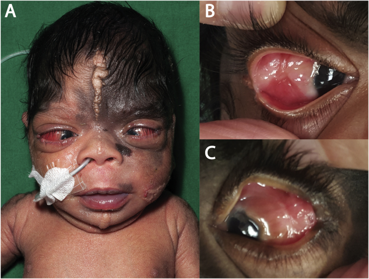
Purpose: To present the ophthalmic manifestations of a 3-month old female with SCALP syndrome.
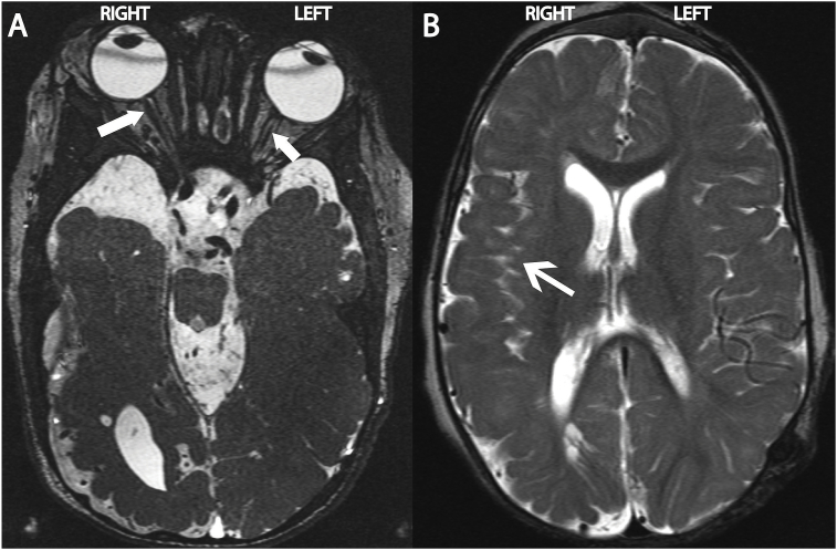
Observations: The patient presented with multiple ocular anomalies including bilateral limbal dermoids, esotropia and left optic nerve hypoplasia.
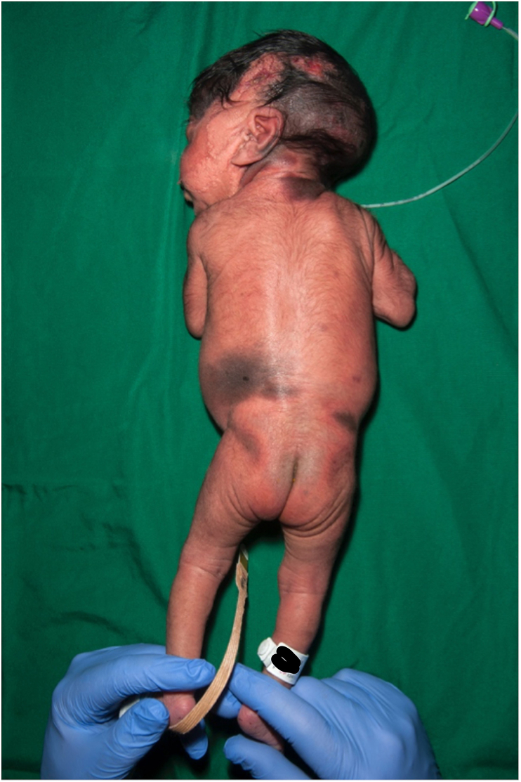
Conclusions: We describe systemic and ocular anomalies in a rare case of SCALP syndrome. This report provides additional information on the ocular anomalies not previously described that may be associated with this clinical entity.
Keywords: Aplasia cutis; Limbal dermoid; Melanocytic naevus; Optic nerve hypoplasia; SCALP syndrome; Sebaceous nevus.
Figures

References
-
- Lam J., Dohil M.A., Eicnhenfield L.F., Cunningham B.B. SCALP syndrome: sebaceous nevus syndrome, CNS malformations, aplasia cutis congenita, limbal dermoid, and pigmented nevus (giant congenital melanocytic nevus) with neurocutaneous melanosis: a distinct syndromic entity. J Am Acad Dermatol. 2008;58:884–888. - PubMed
-
- Baykal C., Buyukabani N., Yazganoglu K.D., Saglik E. Tumors associated with nevus sebaceous. J Dtsch Dermatol Ges. 2006;4:28–31. - PubMed
-
- Mesrati H., Amouri M., Chaaben H., Masmoudi A., Boudaya S., Turki H. Aplasia cutis congenita: report of 22 cases. Int J Dermatol. 2015;54:1370–1375. - PubMed
Publication types
LinkOut - more resources
Full Text Sources
Other Literature Sources
