. 2018 Jul 27;14(7):e1007179.
doi: 10.1371/journal.ppat.1007179. eCollection 2018 Jul.

A cancer-associated Epstein-Barr virus BZLF1 promoter variant enhances lytic infection

Affiliations

A cancer-associated Epstein-Barr virus BZLF1 promoter variant enhances lytic infection

Jillian A Bristol et al. PLoS Pathog. .

Abstract

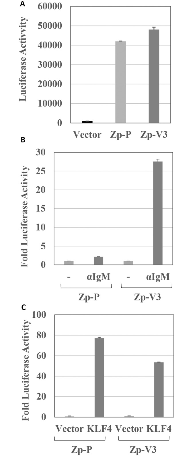
Latent Epstein-Barr virus (EBV) infection contributes to both B-cell and epithelial-cell malignancies. However, whether lytic EBV infection also contributes to tumors is unclear, although the association between malaria infection and Burkitt lymphomas (BLs) may involve excessive lytic EBV replication. A particular variant of the viral promoter (Zp) that controls lytic EBV reactivation is over-represented, relative to its frequency in non-malignant tissue, in EBV-positive nasopharyngeal carcinomas and AIDS-related lymphomas. To date, no functional differences between the prototype Zp (Zp-P) and the cancer-associated variant (Zp-V3) have been identified. Here we show that a single nucleotide difference between the Zp-V3 and Zp-P promoters creates a binding site for the cellular transcription factor, NFATc1, in the Zp-V3 (but not Zp-P) variant, and greatly enhances Zp activity and lytic viral reactivation in response to NFATc1-inducing stimuli such as B-cell receptor activation and ionomycin. Furthermore, we demonstrate that restoring this NFATc1-motif to the Zp-P variant in the context of the intact EBV B95.8 strain genome greatly enhances lytic viral reactivation in response to the NFATc1-activating agent, ionomycin, and this effect is blocked by the NFAT inhibitory agent, cyclosporine, as well as NFATc1 siRNA. We also show that the Zp-V3 variant is over-represented in EBV-positive BLs and gastric cancers, and in EBV-transformed B-cell lines derived from EBV-infected breast milk of Kenyan mothers that had malaria during pregnancy. These results demonstrate that the Zp-V3 enhances EBV lytic reactivation to physiologically-relevant stimuli, and suggest that increased lytic infection may contribute to the increased prevalence of this variant in EBV-associated malignancies.


Conflict of interest statement

The authors have declared that no competing interests exist.

Figures

Fig 1
Fig 1. Zp-V3 is much more responsive to BCR crosslinking than Zp-P.
(A) EBV-negative BJAB cells were transfected with a promoterless luciferase construct, or luciferase constructs driven by the Zp-P or Zp-V3 promoters, and luciferase activity was measured after 24 hours. The amount of luciferase activity for each condition (average and the standard deviation, SD) is shown. (B) BJAB cells were transfected with luciferase constructs driven by the Zp-P or Zp-V3 promoters, and then treated with or without anti-human IgM (10 mg/mL) 24 hours later to activate the B cell receptor (BCR). Luciferase activity was measured at 48 hours after transfection and results were normalized to that each promoter’s untreated condition (set as 1). The fold increase in luciferase activity is shown for each condition (average and SD). (C) EBV-negative NOKs epithelial cells were transfected with the Zp-P or Zp-V3 luciferase vectors, and a KLF4 expression vector or vector control as indicated, and luciferase activity was measured 24 hours post-transfection. The amount of luciferase activity for each condition (average and SD) is shown and results were normalized to that of each promoter’s untreated condition (set as 1).
Fig 2
Fig 2. Residue -141 in the Zp-V3 promoter is critical for response to BCR crosslinking.
(A) The Z promoter sequences in the B95.8 (top) and M81 (bottom) EBV strains are shown. B95.8 is the prototype (Zp-P) while M81 has the 3 polymorphisms at -100, -106, and -141 that define Variant 3 (Zp-V3). There are 4 additional basepair differences between the two Zp sequences, located at -274, -365, -460, and -525 (relative to the transcriptional start site), highlighted in gray. (B) EBV-negative BJAB cells were transfected with wildtype or mutant Zp-V3 luciferase constructs (named to reflect the promoter basepair altered in Zp-V3 relative to the Zp start site) and treated with or without anti-IgM. The luciferase activity for each construct is shown and results were normalized to that of each promoter’s untreated condition (set as 1). (C) BJAB cells were transfected with either the wildtype Zp-P luciferase construct or a mutant Zp-P construct in which the A nucleotide located at -141 is switched to the G nucleotide found in the Zp-V3 sequence. Cells were treated with or without anti-IgM 24 hours after transfection. Luciferase activity was measured at 48 hours after transfection and results were normalized to that of each promoter’s untreated condition (set as 1). The fold increase in luciferase activity is shown for each condition (average and SD).
Fig 3
Fig 3. Zp-V3 (but not Zp-P) binds NFATc1.
(A) EMSA oligonucleotides were designed to encompass the regions of Zp-V3 and Zp-P from -155 to -127, or from -155 to -120 and radiolabeled with 32P. The potential NFAT (in Zp-V3, not Zp-P) and AP1 binding sites (in both variants) are indicated. (B) EBV-negative BJAB cells were transfected with the Zp-V3 luciferase vector, and then treated 24 hours later with or without anti-IgM, in the presence or absence of cyclosporine A (1μM) as indicated. Luciferase activity was measured 24 hours after anti-IgM treatment and results were normalized to that of the Zp-V3 untreated condition. (set as 1). The fold increase in luciferase activity is shown for each condition (average and SD). (C) Nuclear extracts prepared from BJAB cells, treated with or without anti-IgM for 30 minutes or 6 hours, were incubated with the radiolabeled Zp-P or Zp-V3 (-155 to -127) probes in an EMSA. A protein that binds only to the Zp-V3 probe is indicated by an arrow. (D) EMSA was performed using radiolabeled Zp-P and Zp-V3 probes (-155 to -127) and untreated nuclear BJAB extract. Cold competitor DNA containing either the Zp-P or Zp-V3 oligonucleotides, or the consensus binding sites for the transcription factors indicated, was added in some conditions. An arrow depicts bands representing NFAT binding. (E) EMSA was performed using radiolabeled Zp-P and Zp-V3 probes (-155 to -127) and untreated nuclear BJAB extract. In some conditions, antibodies against NFATc1 or C/EBPα were added to the nuclear extract (prior to the addition of the labeled probe) as shown. An arrow depicts bands representing NFAT binding.
Fig 4
Fig 4. The longer Zp-V3 probe contains a cooperative NFAT/AP1 binding motif.
EBV-negative BJAB nuclear extracts, with or without exposure to 6 hours of anti-IgM, were incubated with radiolabeled probes in EMSA assays. (A) A radiolabeled AP1 consensus probe was incubated with nuclear extract obtained from untreated or anti-IgM treated BJAB cells and EMSA performed. Cold competitor DNA containing consensus binding sites for the AP1 or NFAT transcription factors was added in some conditions. (B) Radiolabeled probes containing the Zp-V3 sequences (either from -155 to -127, or from -155 to -120 as indicated) were incubated with nuclear extract obtained from untreated or anti-IgM treated BJAB cells and EMSA performed. Cold competitor DNA containing consensus binding sites for the AP1 or NFAT transcription factors was added in some conditions. Arrows depict bands representing NFAT binding alone and NFAT plus AP1 binding. (C) A radiolabeled probe containing the Zp-V3 sequence from -155 to -120 was incubated with nuclear extract obtained from untreated or anti-IgM treated BJAB cells and EMSA performed. In some conditions, antibodies against cFos or XBP1 were added to the nuclear extract (prior to the addition of the labeled probe) as shown. Arrows depict bands representing NFAT binding alone, NFAT plus AP1 binding, and the NFAT plus AP1 band that is supershifted by the cFos antibody.
Fig 5
Fig 5. NFATc1 and cFos cooperate to activate the Zp-V3.
(A) EBV-negative BJAB cells were transfected with the wildtype Zp-V3 luciferase vector, the Zp-V3-141A, or the Zp-V3 ZIIIA mutant promoter luciferase constructs, with or without plasmids expressing cFos and/or NFATc1 as indicated. The fold increase in luciferase activity induced by co-transfection with cFos or NFATc1 vectors for each promoter construct (relative to the vector control, set as 1) is shown. Error bars indicate standard deviation. (B) Levels of transfected NFATc1 in each condition were determined by immunoblot.
Fig 6
Fig 6. LMP2A, a BCR mimic, activates the Zp-V3 via the NFAT and ZIIIA binding sites.
(A) EBV-negative BJAB cells were transfected with the Zp-P or Zp-V3 luciferase vector with or without a plasmid expressing LMP2A. The fold increase in luciferase activity induced by co-transfection with the LMP2A vector for each promoter construct (relative to the untreated vector control, set as 1) is shown. Error bars indicate standard deviation. (B) BJAB cells were transfected with wildtype Zp-V3 luciferase vector, the Zp-V3-141A, or the Zp-V3 ZIIIA mutant promoter luciferase constructs, with or without a plasmid expressing LMP2A (in the presence or absence of cyclosporine A treatment) as indicated. The fold increase in luciferase activity induced by co-transfection with the LMP2A vector for each promoter construct (relative to the untreated vector control, set as 1) is shown.
Fig 7
Fig 7. Altering the Zp-P -141 nucleotide to that of the Zp-V3 nucleotide in the context of the intact B95.8 viral genome increases lytic protein expression in newly infected B cells.
The -141 residue in the Zp-P of the B95.8 2089 bacmid (encoding GFP) was altered to that of Zp-V3, and infectious viral particles were produced as described in the methods. A revertant construct (Rev), in which the Zp-V3–141 nucleotide was switched back to that of Zp-P, was also constructed. (A) Equal titers of wildtype (WT) B95.8, or the Zp -141G mutant, were used to infect EBV-negative BJAB cells (left panel) or EBV-negative Akata cells (right panel). Immunoblots were performed at the time points indicated after infection to detect the EBV protein EBNA2 (as a control for equal amount of EBV infection), Z, and actin (loading control). (B) Normal primary peripheral B cells were infected with the Zp mutant or revertant virus as indicated and harvested after three days. In the top panel extracts from unsorted cells were immunoblotted for Z, R (BRLF1), and loading control tubulin. The middle panel quantifies the results of the top panel using Image Studio Lite software to normalize the levels of Z and R expression relative to tubulin expression in each cell type. Results are presented as the ratio of Z and R protein in cells infected with the Zp-141G virus relative to Z and R expression in cells infected with the revertant (wildtype) virus. In the bottom panel, the percent GFP+ cells and mean fluorescence intensity of the cells on day 3 (measured by flow cytometry) is shown.
Fig 8
Fig 8. Converting the -141 Zp nucleotide in the intact B95.8 genome to the Zp-V3 nucleotide increases lytic protein expression in stably infected Burkitt cells.
(A) EBV-negative Mutu B cells were infected with wildtype, Zp mutant, or revertant B95.8 (2089) viruses as indicated, and stably selected with hygromycin B for two months. Two different independently selected lines for each virus were then treated for two days with or without ionomycin (in the presence or absence of cyclosporine), and immunoblots were performed to detect EBNA1, EBNA2, LMP1, Z, BMRF1 (early lytic protein), p18 (late lytic protein), and actin. Kem III cell extract was included as a positive control for EBNA1, EBNA2, and LMP1. (B) Mutu cell lines containing Wt or Zp mutant viruses were nucleofected with control siRNA or NFATc1 siRNA. Ionomycin or DMSO control was added after 48 hours, and cells harvested 72 hours post-infection. Immunoblots were performed to detect NFATc1, R, BMRF1, Z, and tubulin (loading control). (C) Mutu cell lines containing Wt or Zp mutant viruses (or mock infected cells) were treated with or without anti-IgG for two days and immunoblots performed to detect BMRF1, Z, and actin (loading control). (D) Mutu cell lines containing Wt, Zp mutant, or revertant viruses were treated with or without TPA plus sodium butyrate (NaBut) (in the presence or absence of cyclosporine) for two days and immunoblots performed to detect Z expression and GAPDH (loading control). (E) ChIP assays were performed using Mutu cell lines containing Wt or Zp mutant viruses treated for three hours with ionomycin. Formaldehyde-fixed cell extracts were immunoprecipitated with control anti-IgG or NFATc1 antibody. qPCR using primers for the EBV Z promoter was performed; results shown are expressed as the amount of Zp complexed to NFATc1 ab relative to the control IgG ab. Data represent three independent experiments.
Fig 9
Fig 9. The Zp-V3 B95.8 mutant is not defective for transforming primary B cells.
Primary peripheral B cells were infected with wildtype, Zp-141G mutant, and revertant virus (MOI 0.25) and maintained for 21 days. 10/10 wells had colony outgrowth for each virus. Representative bright field images from each strain are shown.

References

    1. Hurley EA, Thorley-Lawson DA. B cell activation and the establishment of Epstein-Barr virus latency. J Exp Med. 1988;168: 2059–2075. 10.1084/jem.168.6.2059 - DOI - PMC - PubMed
    1. Decker LL, Klaman LD, Thorley-Lawson DA. Detection of the latent form of Epstein-Barr virus DNA in the peripheral blood of healthy individuals. J Virol. 1996;70: 3286–3289. - PMC - PubMed
    1. Babcock GJ, Decker LL, Volk M, Thorley-Lawson DA. EBV persistence in memory B cells in vivo. Immunity. 1998;9: 395–404. - PubMed
    1. Sixbey JW, Nedrud JG, Raab-Traub N, Hanes RA, Pagano JS. Epstein-Barr virus replication in oropharyngeal epithelial cells. N Engl J Med. 1984;310: 1225–1230. 10.1056/NEJM198405103101905 - DOI - PubMed
    1. Takada K. Cross-linking of cell surface immunoglobulins induces epstein-barr virus in burkitt lymphoma lines. Int J Cancer. 1984;33: 27–32. 10.1002/ijc.2910330106 - DOI - PubMed

Publication types

MeSH terms

Substances