Review
. 2018 Oct:461:23-29.
doi: 10.1016/j.jim.2018.07.007. Epub 2018 Jul 24.

Biosensor-based epitope mapping of antibodies targeting the hemagglutinin and neuraminidase of influenza A virus

Affiliations
Review

Biosensor-based epitope mapping of antibodies targeting the hemagglutinin and neuraminidase of influenza A virus

Zhu Guo et al. J Immunol Methods. 2018 Oct.

Abstract

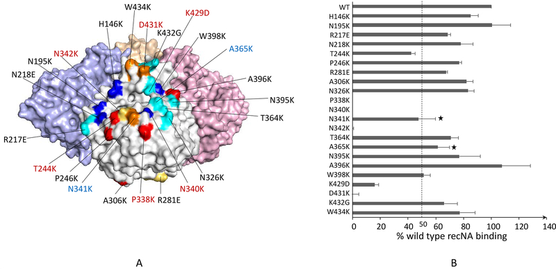
Characterization of the epitopes on antigen recognized by monoclonal antibodies (mAb) is useful for the development of therapeutic antibodies, diagnostic tools, and vaccines. Epitope mapping also provides functional information for sequence-based repertoire analysis of antibody response to pathogen infection and/or vaccination. However, development of mapping strategies has lagged behind mAb discovery. We have developed a site-directed mutagenesis approach that can be used in conjunction with bio-layer interferometry (BLI) biosensors to map mAb epitopes. By generating a panel of single point mutants in the recombinant hemagglutinin (HA) and neuraminidase (NA) proteins of influenza A viruses, we have characterized the epitopes of hundreds of mAbs targeting the H1 and H3 subtypes of HA and the N9 subtype of NA.


Figures

Figure 1.
Figure 1.. A timeline for generation of recombinant HA/NA panels for epitope mapping.
Figure 2.
Figure 2.. Epitope mapping of recombinant mAbs targeting the HA of influenza A(H1N1)pdm09 virus.
A panel of 20 recHAs of A(H1N1)pdm09 virus was produced with each containing a single point mutation. (A) The sequence change and location for each mutant are shown on the 3D structure of trimeric A(H1N1)pdm09 HA (PDB ID 3M6S); mutation G155K is shown twice, once on each monomer. Classical H1 antigenic sites are indicated by different colors on the structure surface: Sa (red), Sb (magenta), Ca (cyan), and Cb (blue). Mutations not overlapping with the classical antigenic sites are indicated in golden yellow color; (B) Mutations that caused significant reductions (≥50%) in mAb binding are indicated on the HA 3D structure. For simplicity, only control antibodies with their corresponding epitopes are signified.
Figure 3.
Figure 3.. Epitope mapping of a broadly neutralizing antibody targeting the H3 HA of influenza A virus.
The mAb F045–092 was produced by co-transfection of its heavy and light chain expression vectors in 293T cells, purified and tested against the H3 HA panel. (A) The sequence changes and locations of mutations as part of the epitope are shown on the 3D structure of trimeric H3 HA (adapted from PDB ID 4WE8). Mutations that caused significant reductions (≥50%) in mAb binding are highlighted in red. Percentage of the Ab binding to the mutants compared to wild type is indicated in the brackets. The overlapped H3 antigenic sites are indicated in different colors: Site A (red), Site B (blue), and Site D (cyan). (B) Association/disassociation curves of BLI for the F045–092 binding to the wild type and mutants with increased off-rate. The biosensors loaded with recHAs were incubated with the Ab for 300 sec at the association step followed by incubation in buffer for 180 sec at the disassociation step as shown on the X-axis. The changes of thickness at the tip of biosensors caused by Ab-Ag binding are shown by the Y-axis. The binding curve for each recHA is labeled with a unique color.
Figure 4.
Figure 4.. Epitope mapping of a mAb with therapeutic potential targeting the N9 NA of influenza A(H7N9) virus.
(A) The sequence changes and locations of the N9 panel mutants are shown on the 3D structure of tetrameric N9 NA (PDB ID 4WMJ). Mutations that caused ≥50% reduction in the mAb 3c10–3 binding are highlighted in red, and increased off-rate in blue. The panel is primarily based on previously identified epitopes of NA as indicated: N1 (red) (Wan et al., 2013), N2 (blue) (Lentz et al., 1984; Webster et al., 1984; Gulati et al., 2002), N8 (yellow) (Saito et al., 1994), and N9 (cyan) (Air et al., 1990; Tulip et al., 1992a; Tulip et al., 1992b). Unique sites selected by us are indicated in golden yellow. (B) Percent response of 3c10–3 binding to each mutant NA compared to the wild type was determined. A ≥50% reduction in binding activity was the cutoff for significance. “*” indicates mutations that caused increased off-rate but less than 50% reduction in binding.

References

    1. Abbott WM, Damschroder MM and Lowe DC, 2014, Current approaches to fine mapping of antigen-antibody interactions. Immunology 142, 526–35. - PMC - PubMed
    1. Abdiche YN, Lindquist KC, Stone DM, Rajpal A and Pons J, 2012, Label-free epitope binning assays of monoclonal antibodies enable the identification of antigen heterogeneity. J Immunol Methods 382, 101–16. - PubMed
    1. Abdiche YN, Miles A, Eckman J, Foletti D, Van Blarcom TJ, Yeung YA, Pons J and Rajpal A, 2014, High-throughput epitope binning assays on label-free array-based biosensors can yield exquisite epitope discrimination that facilitates the selection of monoclonal antibodies with functional activity. PLoS One 9, e92451. - PMC - PubMed
    1. Air GM, 2012, Influenza neuraminidase. Influenza Other Respir Viruses 6, 245–56. - PMC - PubMed
    1. Air GM, Laver WG and Webster RG, 1990, Mechanism of antigenic variation in an individual epitope on influenza virus N9 neuraminidase. Journal of virology 64, 5797–803. - PMC - PubMed

Publication types

MeSH terms

Substances

LinkOut - more resources