Review
. 2018 Sep;43(9):668-684.
doi: 10.1016/j.tibs.2018.06.011. Epub 2018 Jul 24.

How Macrolide Antibiotics Work

Affiliations
Review

How Macrolide Antibiotics Work

Nora Vázquez-Laslop et al. Trends Biochem Sci. 2018 Sep.

Abstract

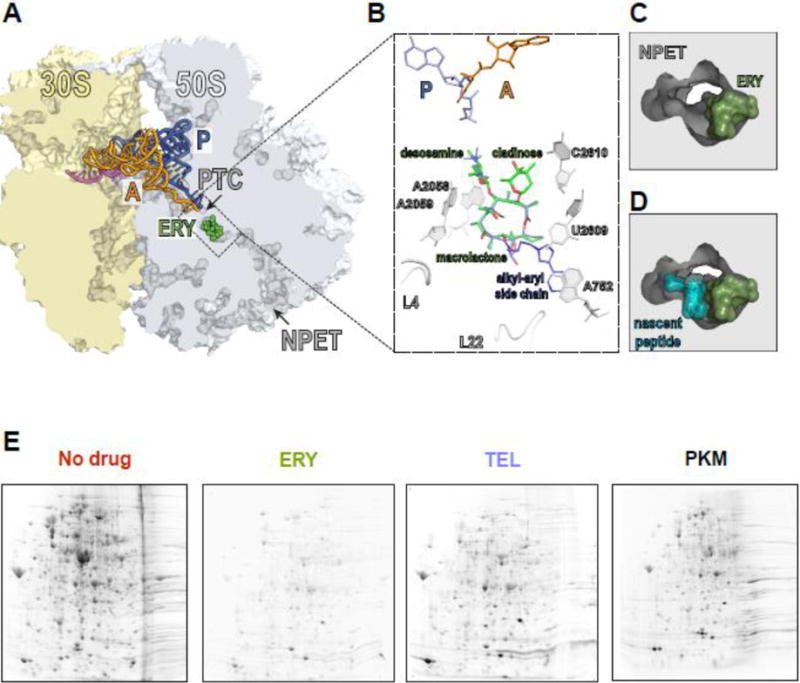
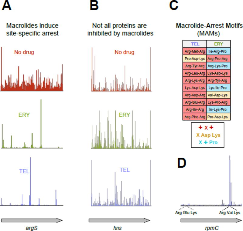
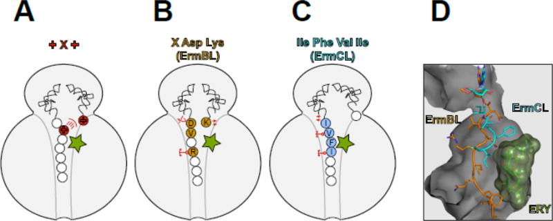
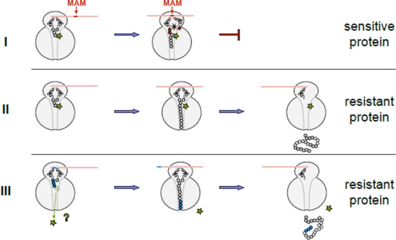
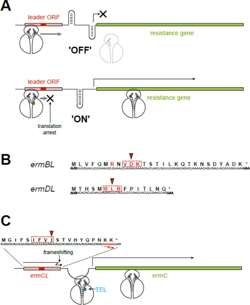
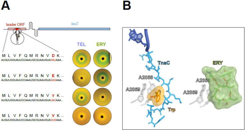
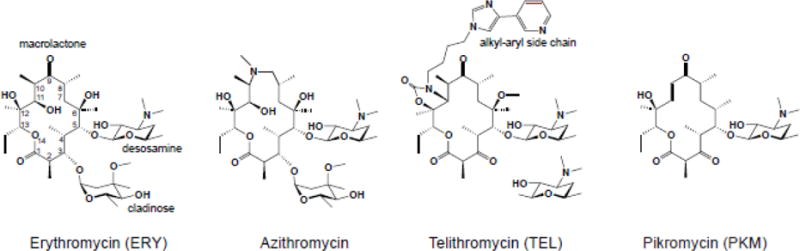
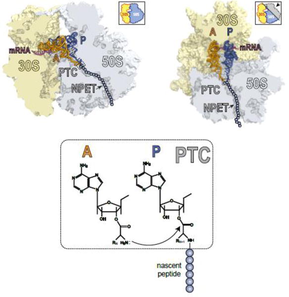
Macrolide antibiotics inhibit protein synthesis by targeting the bacterial ribosome. They bind at the nascent peptide exit tunnel and partially occlude it. Thus, macrolides have been viewed as 'tunnel plugs' that stop the synthesis of every protein. More recent evidence, however, demonstrates that macrolides selectively inhibit the translation of a subset of cellular proteins, and that their action crucially depends on the nascent protein sequence and on the antibiotic structure. Therefore, macrolides emerge as modulators of translation rather than as global inhibitors of protein synthesis. The context-specific action of macrolides is the basis for regulating the expression of resistance genes. Understanding the details of the mechanism of macrolide action may inform rational design of new drugs and unveil important principles of translation regulation.

Keywords: antibiotic; ketolide; macrolide; resistance; ribosome; translation.


Figures

Figure 1
Figure 1. Macrolides narrow the nascent peptide exit tunnel but allow synthesis of certain proteins
(A) The macrolide binding site in the bacterial ribosome. A cross-cut of the ribosome showing the A- and P-site tRNAs (orange and blue, respectively) and a segment of mRNA (magenta). ERY (green) and all the other macrolides bind in the NPET at a short distance from the PTC. (B) The macrolide binding site is composed primarily of rRNA. The macrolactone ring of different macrolides (for comparison, the structures of ERY, green, and TEL, blue, have been superimposed) lays flat against the NPET wall and the C3 and C5 sugars protrude towards the PTC but do not reach its active site. C5 desosamine interacts with the splayed-out A2058 and A2059 rRNA residues in the NPET. C2610 nucleotide contacts C3 cladinose (present in ERY, but lacking in TEL). The alkyl-aryl side chain of ketolides, such as TEL, usually extends away from the PTC: In E. coli, it interacts with the A752-U2609 base pair, but its placement may differ in other bacteria [, Dunkle, 2010 #7182, 75]. The loops of proteins L4 and L22, which form a constriction in the NPET, may directly interact with the side chains of some macrolides [76]. (C) and (D) View into the NPET from the PTC showing that the macrolide (ERY) narrows the tunnel’s aperture (C). The remaining opening of the NPET is nevertheless wide enough for a nascent peptide to be threaded through (D). (E) Specific proteins are synthesized in macrolide-treated cells. Gel electrophoresis analysis of radiolabeled proteins translated in E. coli cells in the absence of antibiotics (No drug), or exposed to high concentrations of ERY, semi-synthetic ketolide TEL, or the natural ketolide pikromycin (PKM).
Figure 2
Figure 2. Macrolides arrest the ribosome at specific mRNA codons
(A) Treatment of E. coli cells with high concentrations of ERY or TEL leads to dramatic redistribution of ribosomes along the genes (illustrated by the ribosome densities in the representative gene argS). The discrete peaks of ribosome density at specific codons of argS in the ERY- or TEL-treated cells reflect context-specific translation arrest [24, 25]. (B) Some ORFs are completely translated in macrolide-treated cells. The occurrence of ribosome density throughout the representative gene hns in cells treated with TEL reveals HN-S as one of the proteins fully translated in the presence of this antibiotic (See Figure 1D). (C) Ribosome with bound macrolide struggles to polymerize specific sequences, the Macrolide Problematic Motifs (MAMs). The table lists the 10 most prevalent MAMs enriched in the sites of translation arrest in the TEL- and ERY-treated cells [25]. The consensus sequences of the common MAMs are listed underneath the table (‘X’ indicates any amino acid, ‘+’ indicates Arg or Lys). (D) Translation arrest at an MAM can be influenced by a more extended context, likely involving other segments of the nascent protein chain. The shown example illustrates how TEL-bound ribosomes could easily translate through the usually problematic Arg-Glu-Lys sequence (an MAM of the + X + type) present within the early codons of rpmC but became arrested at the second +X+ MAM (Arg-Val-Lys) located towards the end of the ORF.
Figure 3
Figure 3. Macrolides are selective modulators of the peptidyl transferase center
The interplay between the macrolide molecule and a MAM-containing nascent peptide alters the PTC properties and inhibits peptide bond formation. In (A–C), the residues critical for stalling are colored and in (B) and (C) are indicated with the single-letter code. (A) Stalling at the +X+ MAM may occur because the macrolide orients the lengthy, positively charged side chain of the penultimate amino acid of the nascent peptide towards the PTC A site, preventing the accommodation of the similarly long and positively charged acceptor amino acid. (B) The macrolide imposes an unfavorable orientation of the C-terminal Asp residue of the ErmBL peptide that contains the X-Asp-Lys MAM [19, 21]. The placement of the acceptor Lys in the A site is also suboptimal. The orientation of several key PTC nucleotides is altered in the stalled ribosome. The mobility of the nascent peptide in the NPET is restricted due to the antibiotic presence and specific interactions of the Arg residue of the nascent chain with rRNA of the tunnel wall. (C) Interactions of the ErmCL nascent chain with the NPET nucleotides and antibiotic misplace the peptide’s C-terminal residue in the PTC [20]. The adverse conformation of the PTC nucleotides prevents accommodation of the A site amino acid [20, 72]. (D). The different placement of the ErmBL and ErmCL nascent peptides in the NPET of the ERY-stalled ribosome shows that the peptide trajectory depends on the amino acid sequence.
Figure 4
Figure 4. The general model of macrolide action upon protein synthesis
Translation of any protein can be initiated by the macrolide-bound ribosome and the N-termini of the majority of polypeptides can be threaded through the drug-obstructed NPET. The subsequent fate of the protein being made depends on its sequence and the structure of the bound antibiotic. (I) Translation of sensitive proteins is interrupted because the drug-bound ribosome is unable to efficiently catalyze peptide bond formation during synthesis of the MAM sequence. The structure of the macrolide molecule bound in the NPET dictates the spectrum of the MAM sequences and therefore, defines which proteins will be inhibited. If translation is arrested close to the start of the ORF, when the nascent peptide is short (<10 amino acids), peptidyl-tRNA likely dissociates from the ribosome (the effect known as peptidyl-tRNA drop-off) [7, 8, 77]. (II) If the protein sequence lacks MAMs, its translation will proceed unimpeded and the full-size protein will be produced in the cell exposed to the antibiotic. (III) Hypothetically, some proteins might be able to dislodge the antibiotic from the ribosome at the early stages of translation; in this case, the drug-free ribosome completes the synthesis of such protein. Antibiotic eviction has been observed with some artificial short peptides [–16], but has not been demonstrated yet for the cellular polypeptides; yet this scenario remains a possibility for at least some of the bacterial proteins.
Figure 5
Figure 5. Inducible resistance exploits the context specific action of macrolides
(A) In an inducible macrolide resistance operon, the resistance gene is preceded by a regulatory leader ORF. In the absence of antibiotic, the leader ORF is translated while the resistance gene is not expressed, because the ‘off’ conformation of the intergenic region of mRNA precludes the access to its translation initiation site. When macrolide is present, translation of the leader ORF is arrested at a specific codon within an MAM (red rectangle). The stalled ribosome re-arranges the mRNA structure into the ‘on’ conformation, releasing the initiation site of the resistance gene and activating its expression. The MAM location is optimal for the paused ribosome to activate the isomerization of the mRNA structure. In a similar scenario, ribosome stalling at the leader ORF can activate attenuated transcription of some of the resistance genes. (B) Programmed ribosome arrest relies on the MAMs (dotted rectangle) encoded in the leader ORFs. The Val-Asp-Lys (the X-Asp-Lys MAM) is embedded in the ermBL gene, while the Arg-Lys-Arg (the +X+ MAM), is found in ermDL. The codon where the ribosome stalls in the presence of antibiotic is indicated by an arrowhead. The amino acids essential for programmed translation arrest are shown in red. (C) Macrolide-induced miscoding accounts for an unorthodox induction of resistance. The ribosome with bound TEL (blue star) ignores the stall site within the ermCL ORF, where ERY would arrest translation at the Ile-9 codon (red arrowhead) of the Ile-Phe-Val-Ile MAM (dotted rectangle). Therefore, the TEL-bound ribosome traverses the entire ermCL ORF and reaches its last two Lys codons with the sequence AAA AAA (red) where TEL stimulates (−1) ribosomal frameshifting. Upon frameshifting, the 0-frame stop codon of the ermCL ORF is skipped and the ribosome continues translation through the intergenic region, dynamically unfolding the mRNA structure and releasing the translation initiation site of the resistance gene.
Figure 6
Figure 6. Nascent peptides turn the ribosome into a small molecule sensor
(A) Single amino acid changes in the ErmBL nascent peptide alters the ribosomal response to structurally different macrolides. The reporter cassette mimics an inducible erm resistance operon (see Figure 5), in which the resistance gene was replaced with lacZ. The reporter induction is visualized by the green color of the cell lawn in the vicinity of an antibiotic-containing disk placed on the agar plate. Both ketolide TEL or cladinose-containing ERY arrest ermBL translation and activate the reporter when the 10 th codon (red) of ermBL specifies Asp (wild type). Changing th the 10 codon to Glu preserves the response to ERY but eliminates the response to TEL. Tyr-10 allows the ribosome to respond exclusively to ketolides. Val-10 precludes the response to either TEL or ERY. (B) The TnaC nascent peptide allows sensing of tryptophan. Activation of the tna operon depends on ribosome stalling on the tnaC leader ORF [60, 61]. Structural studies of the TnaC-stalled ribosome suggest that one of the two observed tryptophan molecules binds at the A2058/A2059 crevice (left image) [63], the same site that is exploited by macrolide antibiotics for binding to the ribosome (right image) [78]. Similar to the coordinated action of macrolides and nascent peptide in inducing translation arrest, the TnaC peptide cooperates with tryptophan to disrupt the PTC function and stall the ribosome at the last sense codon of the tnaC ORF.
Box figure I
Box figure I
Box figure II
Box figure II

References

    1. Wilson DN. Ribosome-targeting antibiotics and mechanisms of bacterial resistance. Nat Rev Microbiol. 2014;12:35–48. - PubMed
    1. Lin J, et al. Ribosome-targeting antibiotics: modes of action, mechanisms of resistance, and implications for drug design. Annu Rev Biochem. 2018;87:18.1–18.28. - PMC - PubMed
    1. Contreras A, Vazquez D. Cooperative and antagonistic interactions of peptidyl-tRNA and antibiotics with bacterial ribosomes. Eur J Biochem. 1977;74:539–547. - PubMed
    1. Odom OW, et al. The synthesis of polyphenylalanine on ribosomes to which erythromycin is bound. Eur J Biochem. 1991;198:713–722. - PubMed
    1. Arevalo MA, et al. Protein components of the erythromycin binding site in bacterial ribosomes. J Biol Chem. 1988;263:58–63. - PubMed

Publication types

MeSH terms