Review
. 2019 Oct;60(10):1720-1732.
doi: 10.1194/jlr.R087510. Epub 2018 Jul 27.

Muscle and adipose tissue insulin resistance: malady without mechanism?

Affiliations
Review

Muscle and adipose tissue insulin resistance: malady without mechanism?

Daniel J Fazakerley et al. J Lipid Res. 2019 Oct.

Abstract

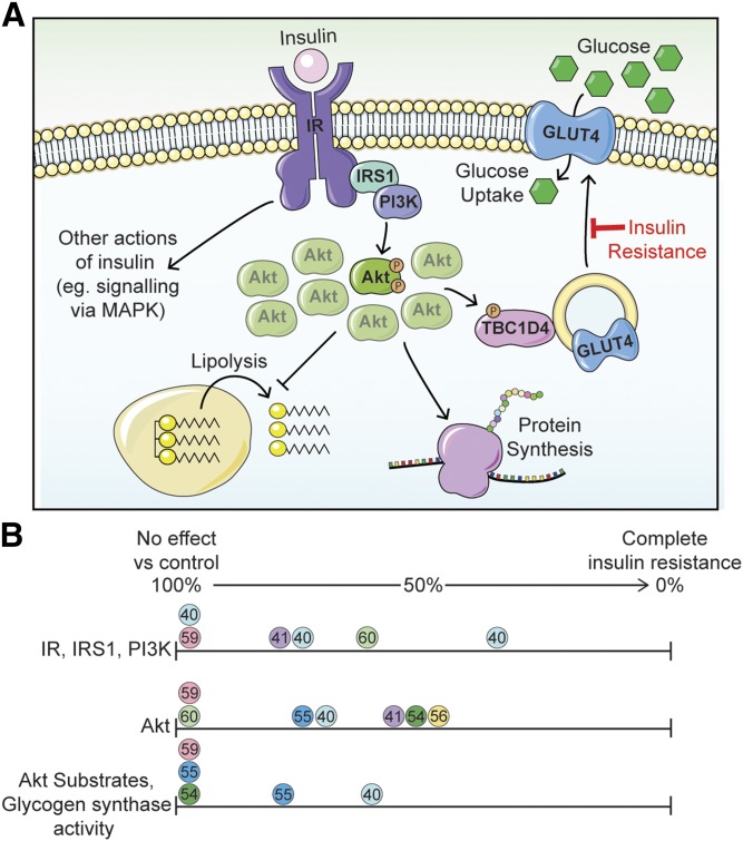
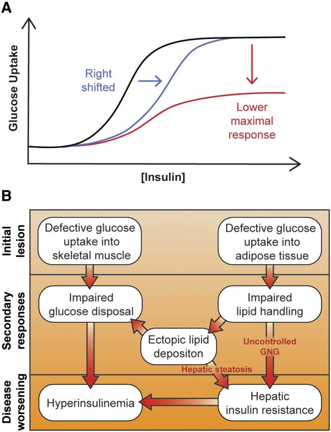
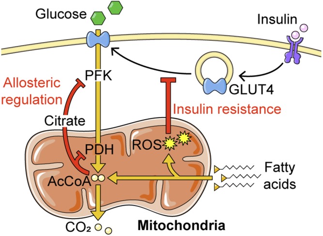
Insulin resistance is a major risk factor for numerous diseases, including type 2 diabetes and cardiovascular disease. These disorders have dramatically increased in incidence with modern life, suggesting that excess nutrients and obesity are major causes of "common" insulin resistance. Despite considerable effort, the mechanisms that contribute to common insulin resistance are not resolved. There is universal agreement that extracellular perturbations, such as nutrient excess, hyperinsulinemia, glucocorticoids, or inflammation, trigger intracellular stress in key metabolic target tissues, such as muscle and adipose tissue, and this impairs the ability of insulin to initiate its normal metabolic actions in these cells. Here, we present evidence that the impairment in insulin action is independent of proximal elements of the insulin signaling pathway and is likely specific to the glucoregulatory branch of insulin signaling. We propose that many intracellular stress pathways act in concert to increase mitochondrial reactive oxygen species to trigger insulin resistance. We speculate that this may be a physiological pathway to conserve glucose during specific states, such as fasting, and that, in the presence of chronic nutrient excess, this pathway ultimately leads to disease. This review highlights key points in this pathway that require further research effort.

Keywords: glucose transporter type 4; insulin signaling; mitochondria; oxidants.


Figures

Fig. 1.
Fig. 1.
Insulin resistance is not due to a generalized impairment in insulin signaling. A: Insulin stimulates a signaling cascade via Akt kinase to regulate biological processes, such as lipolysis, protein synthesis, and glucose transport, the latter via GLUT4 translocation to the plasma membrane. See the main text for additional details. Only a small percentage of the total phosphorylatable pool of Akt is required to maximally regulate downstream biological processes. Insulin-regulated glucose transport is selectively impaired in common insulin resistance, while other insulin-regulated processes in the same cell are less affected. We refer to this as cis selective insulin resistance. B: Analysis of studies examining insulin signaling in the muscles of insulin-resistant subjects that underwent a hyperinsulinemic-euglycemic clamp. The phosphorylation or activity of signaling elements upstream of Akt (IR, IRS1, PI3K), at Akt (Akt), and downstream of Akt (Akt substrates, glycogen synthase) are expressed as a percentage of values from healthy control subjects. Circles of the same color represent data within a single study; reference numbers are included within the circles. References (54) and (60) (green circles) highlight that defects earlier in the pathway are often not translated downstream.
Fig. 2.
Fig. 2.
Impaired glucose transport at the center of common insulin resistance. A: Theoretical insulin dose response curves for glucose uptake in human subjects. A reduction in signal transmission via proximal signaling components (e.g., IR, IRS1, PI3K, Akt) would cause this curve to be right-shifted (blue line), requiring a higher insulin dose to achieve the same output. Alternatively, a “post-signaling” impairment would result in a lower maximal response (red line). Both are observed in common insulin resistance (45). B: Model for how initial reductions in glucose uptake into adipose and muscle tissue can precipitate hepatic and whole-body insulin resistance via impaired glucose disposal and lipid handling. Details are in the main text.
Fig. 3.
Fig. 3.
ROS mediate the interplay between fatty acid oxidation and GLUT4 translocation. ROS arising from fatty acid oxidation impair insulin-stimulated GLUT4 translocation to inhibit glucose uptake (insulin resistance) by an unknown mechanism. This complements the well-established mechanism by which fatty acid oxidation inhibits glucose metabolism, via targeting phosphofructokinase (PFK) and pyruvate dehydrogenase (PDH) by allosteric regulation.

References

    1. Abdul-Ghani M. A., Tripathy D., and DeFronzo R. A.. 2006. Contributions of beta-cell dysfunction and insulin resistance to the pathogenesis of impaired glucose tolerance and impaired fasting glucose. Diabetes Care. 29: 1130–1139. - PubMed
    1. Meyer C., Pimenta W., Woerle H. J., Van Haeften T., Szoke E., Mitrakou A., and Gerich J.. 2006. Different mechanisms for impaired fasting glucose and impaired postprandial glucose tolerance in humans. Diabetes Care. 29: 1909–1914. - PubMed
    1. Chen D. L., Liess C., Poljak A., Xu A., Zhang J., Thoma C., Trenell M., Milner B., Jenkins A. B., Chisholm D. J., et al. . 2015. Phenotypic characterization of insulin-resistant and insulin-sensitive obesity. J. Clin. Endocrinol. Metab. 100: 4082–4091. - PubMed
    1. Samuel V. T., and Shulman G. I.. 2016. The pathogenesis of insulin resistance: integrating signaling pathways and substrate flux. J. Clin. Invest. 126: 12–22. - PMC - PubMed
    1. Petersen M. C., and Shulman G. I.. 2017. Roles of diacylglycerols and ceramides in hepatic insulin resistance. Trends Pharmacol. Sci. 38: 649–665. - PMC - PubMed

LinkOut - more resources