. 2018 Jul 27;9(1):2951.
doi: 10.1038/s41467-018-05346-7.

Macrophages are exploited from an innate wound healing response to facilitate cancer metastasis

Affiliations

Macrophages are exploited from an innate wound healing response to facilitate cancer metastasis

Tamara Muliaditan et al. Nat Commun. .

Abstract

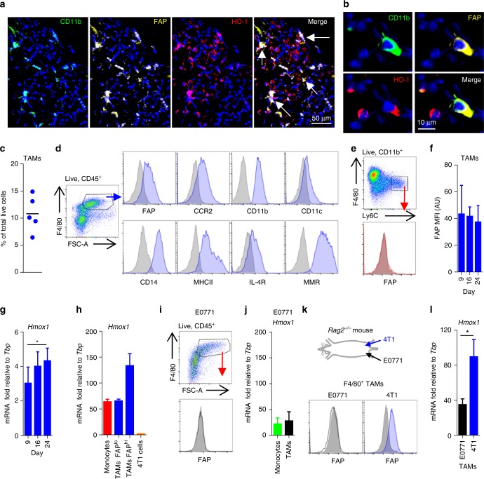
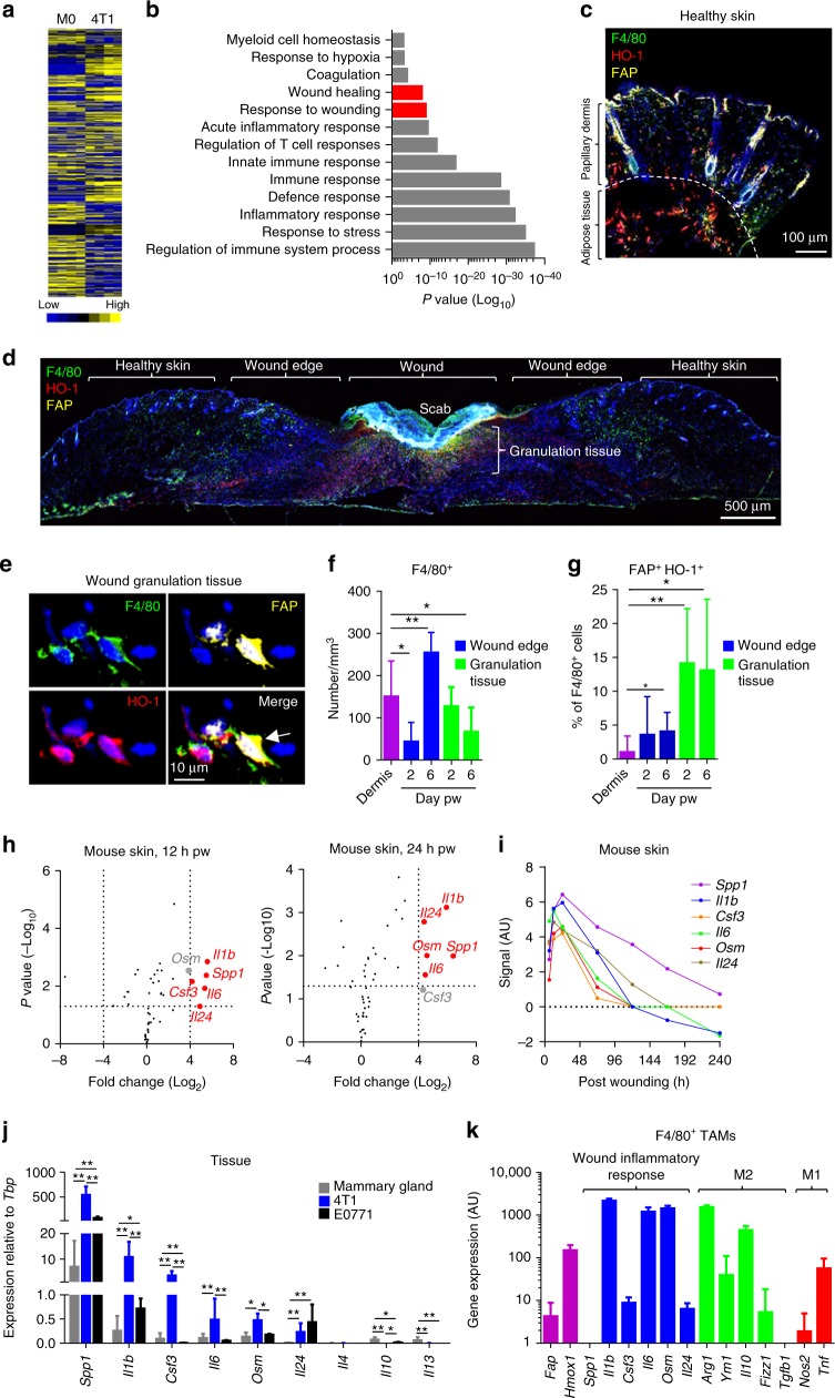
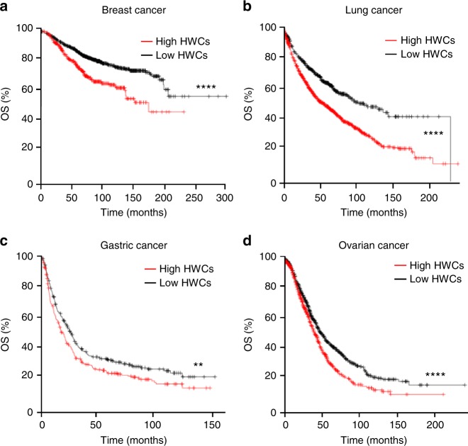
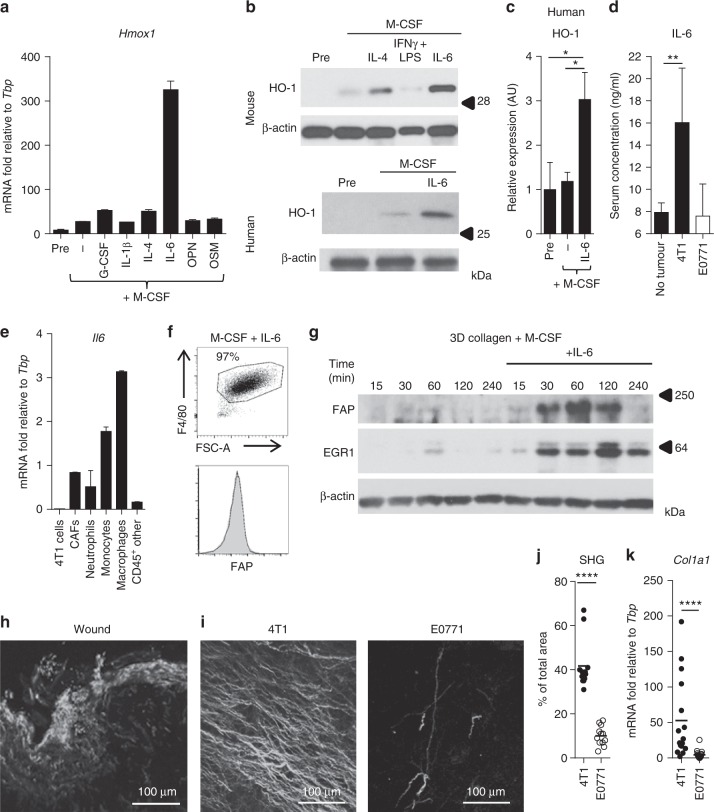
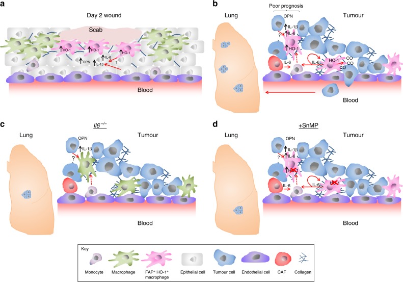
Tumour-associated macrophages (TAMs) play an important role in tumour progression, which is facilitated by their ability to respond to environmental cues. Here we report, using murine models of breast cancer, that TAMs expressing fibroblast activation protein alpha (FAP) and haem oxygenase-1 (HO-1), which are also found in human breast cancer, represent a macrophage phenotype similar to that observed during the wound healing response. Importantly, the expression of a wound-like cytokine response within the tumour is clinically associated with poor prognosis in a variety of cancers. We show that co-expression of FAP and HO-1 in macrophages results from an innate early regenerative response driven by IL-6, which both directly regulates HO-1 expression and licenses FAP expression in a skin-like collagen-rich environment. We show that tumours can exploit this response to facilitate transendothelial migration and metastatic spread of the disease, which can be pharmacologically targeted using a clinically relevant HO-1 inhibitor.


Conflict of interest statement

The authors declare no competing interests.

Figures

Fig. 1
Fig. 1
FAP+ HO-1+ TAMs in human and mouse breast adenocarcinoma are a tumour-educated phenotype. a, b Representative images of frozen human invasive ductal mammary carcinoma sections stained with DAPI (nuclei; blue) and antibodies against CD11b (green), FAP (yellow) and HO-1 (red) (representative images from n = 3 tumours). Arrows indicate cells co-expressing CD11b, FAP and HO-1. c, d Abundance (c) and surface characterisation (d) of live (7AAD) CD45+ F4/80+ TAMs from enzyme-dispersed 4T1 tumours (at day 24 post inoculation) as assessed using flow cytometry. Each point in c represents live F4/80+ cells in an individual tumour. Histograms represent positive staining for the markers shown (blue shaded) against that of the respective isotype controls (grey shaded). e Flow cytometry gating strategy for live CD11b+ F4/80low Ly6Chi monocytes in a representative enzyme-dispersed 4T1 tumour; histogram shows surface FAP staining (red shaded) against that of the isotype control (grey shaded). f Median fluorescence intensity (MFI) of FAP surface expression with fluorescence minus one (FMO) background staining subtracted on CD45+ F4/80+ TAMs from enzyme-dispersed 4T1 tumours on the indicated day post injection of the tumour cells (n = 6). g, h Hmox1 mRNA expression relative to the housekeeping gene Tbp in 4T1 tumour tissue on the indicated day post inoculation of the tumour cells (n = 6–8 tumours per time point) (g) and FACS-sorted monocytes, FAPlo F4/80hi TAMs and FAPhi F4/80hi TAMs and 4T1 tumour cells (CD45 Thy1.2 CD31) from duplicate wells from a 4T1 tumour, data representative of duplicate experiments (h). i Representative FAP staining of live (7AAD) CD45+ F4/80hi TAMs from an enzyme-dispersed E0771 tumour as assessed using flow cytometry. Histogram represents positive staining for FAP (black line) against isotype control staining (grey shaded). j Hmox1 mRNA expression relative to the housekeeping gene Tbp in FACS-sorted monocytes (CD11b+ F4/80lo Ly6Chi) and TAMs (F4/80hi) from duplicate wells from an E0771 tumour, data representative of duplicate experiments. k, l 4T1 (blue) and E0771 (black open) tumours were grown concurrently in opposing mammary fat pads in Rag2/ mice. k Schematic representation of the experimental setting (top) and representative FAP expression as assessed using flow cytometry on live CD45+ F4/80hi TAMs from enzyme-dispersed tumours (bottom). l Hmox1 mRNA expression relative to the housekeeping gene Tbp in MACS-sorted TAMs from enzyme-dispersed tumours (n = 4). Bar charts represent mean + s.d. *P< 0.05
Fig. 2
Fig. 2
FAP+ HO-1+ macrophages represent a wound healing response phenotype. a Heat map of raw gene expression values from splenic-derived M-CSF basal macrophages (M0) and 4T1 TAMs (n = 4 per condition). b Gene ontology enrichment analysis of M0 vs. 4T1 TAMs, showing selected pathways of interest that were significantly enriched. ce Skin sections stained using DAPI (nuclei, blue) and antibodies against F4/80 (green), FAP (yellow) and HO-1 (red). c Image of healthy skin taken using a ×40 objective. d Image of wounded skin taken using a ×4 objective 6 days post wounding (pw). e High magnification image of example F4/80+ FAP+ HO-1+ cells found in the granulation tissue; arrow marks cell of interest. f, g Quantification of F4/80+ cells normalised to area (f) and percentage of F4/80+ cells that co-expressed FAP and HO-1 (g) as an average of multiple fields of view in the healthy dermis and in the respective area of the wound environment (n = 6 mice per condition). Statistics were performed to compare each condition to healthy dermis. h Volcano plots showing changes in cytokine gene expression in skin in response to wounding using a 1 mm punch biopsy needle. Genes marked in red represent cytokines most highly induced 12 h (left) and 24 h (right) pw (P 0.05; Log2 (fold change) ≥4). i Expression levels of the indicated genes (h) over the duration of the wound response to a 1 mm punch biopsy needle. j mRNA expression of the indicated cytokine genes relative to the housekeeping gene Tbp in healthy mammary gland (n = 6) and late stage (>20 day post inoculation) 4T1 (blue) and E0771 (black) tumours (n = 6). k M0-subtracted gene expression from 4T1 TAMs, displaying selected genes of interest (n = 4). Bar charts represent mean + s.d. *P< 0.05, **P ≤ 0.01
Fig. 3
Fig. 3
The presence of a healing wound-like cytokine response is associated with poor prognosis in human cancers. Kaplan–Meier survival curves showing overall survival (OS) with high (red) and low (black) tumour expression of HWCs (SPP1, IL1B and IL6) for patients with a breast cancer (n = 996 in HWCslo group and 406 in HWCshi group), b lung cancer (n = 622 in HWCslo group and 1304 in HWCshi group), c gastric cancer (n = 313 in HWCslo group and 280 in HWCshi group) and d ovarian cancer (n = 843 in HWCslo group and 813 in HWCshi group). **P ≤ 0.01, ****P ≤ 0.0001
Fig. 4
Fig. 4
IL-6 and collagen drive FAP+ HO-1+ TAM differentiation in vitro. a Hmox1 mRNA expression relative to the housekeeping gene Tbp in bone marrow-derived (BM) cells before (Pre) and after macrophage differentiation in the presence of indicated cytokines at 50 ng/ml for 72 h (n = 2 wells; representative of three experiments). b Western blot for HO-1 and the loading control β-actin in BM cells before (Pre) and after 72 h culture in the presence of 50 ng/ml M-CSF with/without 50 ng/ml IL-4, IFNγ + LPS or IL-6 (top) and human peripheral blood (PB)-derived macrophages before (Pre) and after 72 h culture in the presence of 50 ng/ml M-CSF with/without 50 ng/ml IL-6 (bottom). c Quantitation of HO-1 expression in human PB-derived macrophages from individual healthy volunteers (n = 5) as described in b. d Serum IL-6 concentrations in tumour-free mice and in those bearing late stage (>20 days post inoculation) 4T1 (black) or E0771 (white) tumours (n = 5) as determined by ELISA. e Representative Il6 mRNA expression relative to Tbp in FACS-sorted tumoural cell populations from a 4T1 tumour (n = 2 wells, representative of duplicate experiments). f Representative gating, purity and FAP surface expression of BM cells cultured for 72 h in the presence of 50 ng/ml M-CSF and IL-6. g Representative western blot analysis for FAP (top), EGR1 (middle) and β-actin (bottom) in BM cells cultured for 72 h with 50 ng/ml M-CSF with/without 50 ng/ml IL-6, and subsequently incubated for indicated times on 3D murine type I collagen. h, i Representative images of second-harmonic generation (SHG) imaging of fibrillar collagen in frozen sections of wounded skin (h) and of 4T1 and E0771 tumours (i). j Quantitation of SHG signal in 4T1 and E0771 tumours (n = 12 tumours (>20 days post inoculation) in each group; average across multiple fields of view). k Col1a1 mRNA expression relative to Tbp in 4T1 (n = 16) and E0771 (n = 18) tumours (>20 days post inoculation). Bar charts are presented as mean + s.d. *P ≤ 0.05, **P ≤ 0.01, ****P ≤ 0.0001
Fig. 5
Fig. 5
IL-6 regulates the FAP+ HO-1+ TAM phenotype in vivo. a Example growth curves of 4T1 tumours grown in WT (blue) or Il6−/− (red) mice (n = 8 per condition). At day 23 post inoculation of 4T1 cells, tumours were excised and analysed. b, c The abundance of live CD45+ F4/80+ TAMs (b) and their median fluorescence intensity (MFI) of FAP surface expression with FMO background staining subtracted (c) (n = 11 WT mice and 7 Il6−/− mice). d Representative frozen sections of 4T1 tumours stained with DAPI (nuclei; blue) and antibodies against F4/80 (green) and HO-1 (red). e Quantification of the fraction of F4/80+ cells co-expressing HO-1 (n = 5 tumours) as shown in d. f Representative frozen sections stained with DAPI (nuclei; blue) and antibodies against F4/80 (green) and EGR1 (red). g Quantification of nuclear EGR1 in F4/80+ cells (n = 20 areas across three tumours per condition). h Col1a1 mRNA expression in 4T1 tumours relative to the housekeeping gene Tbp. Growth curve in a is presented as mean ± s.e.m. and bar charts represent mean + s.d. **P ≤ 0.01, ****P ≤ 0.0001
Fig. 6
Fig. 6
Perivascular FAP+ HO-1+ macrophages can facilitate transendothelial migration of tumour cells through their HO activity. a Representative images of frozen sections of a 4T1 tumour stained with DAPI (nuclei; blue) and antibodies against F4/80 (green) and HO-1 (red); functional vasculature was labelled in vivo using dextran-FITC (yellow). b Quantification of F4/80+ TAMs co-expressing HO-1 in avascular and perivascular regions (n = 12 sections across four tumours in each group). c Tumour growth curves of mice bearing established 4T1 tumours treated with vehicle (blue) or 25 μmol/kg/day SnMP (orange) (n = 6), arrow marks the initiation of treatment. d Metastatic nodules on the lung surface of mice bearing 4T1 tumours treated with vehicle (n = 16) or 25 μmol/kg/day SnMP (n = 15) at day 24 post inoculation of tumour cells (data from three independent experiments presented). ei 4T1 tumours or lungs in mice treated with vehicle (blue) or SnMP (orange) as described in c were analysed. e Abundance of TAMs, monocytes and neutrophils in enzyme-dispersed tumours (n = 5 per condition). f Representative flow cytometry gating strategy for MAMs. g Representative surface FAP staining (blue) and isotype control staining (grey shaded) of MAMs in the lungs of 4T1 tumour-bearing mice (day 24). h, i Abundance of lung neutrophils (h) and MAMs (i) (n = 4 per condition). j Representative flow cytometry gating strategy (left) and abundance (right) of 4T1-eGFP cells in enzyme-digested lungs 48 h after i.v. injection of 4T1-eGFP in mice treated with vehicle (blue) or 25 μmol/kg/day SnMP (orange) (n = 10 mice). km Abundance of metastatic nodules on the surface of lungs in mice bearing 4T1 tumours grown in WT (n = 17) and Il6−/− (n = 9) mice (k), WT (n = 12) and Il4r−/− (n = 8) mice (l), or in syngeneic mice bearing 4T1 (n = 9) or E0771 (n = 9) tumours (m). n Schematic representation of the transendothelial migration assay; EC endothelial cells. o, p Relative transendothelial migration of 4T1-eGFP cells (o) and permeability of the EC layer to albumin (p) in the presence or absence of HO-1+ Ly6Chi monocyte-derived macrophages (M(IL-6)) with/without 25 μM SnMP (orange) (n = 4 wells; representative of duplicate experiments) over a 24 h period. q Relative transendothelial migration of 4T1-eGFP cells in the absence (n = 12) or presence of 20 μM Iron (II) chloride (n = 8), 5 μM biliverdin (n = 8) or 250 ppm CO (n = 3) (pooled data from separate experiments). Bar charts represent mean + s.d. *P < 0.05, ** P < 0.01, ****P < 0.0001
Fig. 7
Fig. 7
Macrophages are exploited from an innate wounding response to facilitate metastasis. a The acute wounding inflammatory response results in a microenvironment with high levels of osteopontin (OPN), IL-1β and IL-6. In response to IL-6, in combination with the collagen-rich environment of the skin, the macrophages adopt an FAP+ HO-1+ wound response phenotype. b Tumours exploit the acute wound healing response of macrophages, by inducing IL-6 expression in these cells, as well as other stromal cells, by a currently unknown mechanism. IL-6 signals to monocytes/macrophages resulting in upregulation of HO-1 expression. The tumour microenvironment is also rich in collagen, which licences FAP expression on TAMs in an IL-6-dependent manner. FAP+ HO-1+ TAMs are predominantly located in the perivascular region and facilitate transendothelial migration of tumour cells through their production of CO, facilitating tumour cell intravasation into the bloodstream and eventual colonisation in the lung. c In the absence of IL-6 (Il6−/− mice), the tumour is compromised in its ability to generate the wound healing response TAM phenotype, and lung metastasis is reduced. d FAP+ HO-1+ TAMs can be therapeutically targeted using SnMP to inhibit their production of CO, which prevents transendothelial migration of the tumour cells and subsequent lung colonisation

References

    1. Noy R, Pollard JW. Tumor-associated macrophages: from mechanisms to therapy. Immunity. 2014;41:49–61. doi: 10.1016/j.immuni.2014.06.010. - DOI - PMC - PubMed
    1. Lin EY, et al. Vascular endothelial growth factor restores delayed tumor progression in tumors depleted of macrophages. Mol. Oncol. 2007;1:288–302. doi: 10.1016/j.molonc.2007.10.003. - DOI - PMC - PubMed
    1. Arnold JN, Magiera L, Kraman M, Fearon DT. Tumoral immune suppression by macrophages expressing fibroblast activation protein alpha and heme oxygenase-1. Cancer Immunol. Res. 2014;2:121–126. doi: 10.1158/2326-6066.CIR-13-0150. - DOI - PMC - PubMed
    1. DeNardo DG, et al. Leukocyte complexity predicts breast cancer survival and functionally regulates response to chemotherapy. Cancer Discov. 2011;1:54–67. doi: 10.1158/2159-8274.CD-10-0028. - DOI - PMC - PubMed
    1. Mitchem JB, et al. Targeting tumor-infiltrating macrophages decreases tumor-initiating cells, relieves immunosuppression, and improves chemotherapeutic responses. Cancer Res. 2013;73:1128–1141. doi: 10.1158/0008-5472.CAN-12-2731. - DOI - PMC - PubMed

Publication types

MeSH terms

LinkOut - more resources