. 2018 Jul 27;8(1):11339.
doi: 10.1038/s41598-018-29477-5.

Immunophenotyping and transcriptional profiling of in vitro cultured human adipose tissue derived stem cells

Affiliations

Immunophenotyping and transcriptional profiling of in vitro cultured human adipose tissue derived stem cells

Alina Mieczkowska et al. Sci Rep. .

Abstract

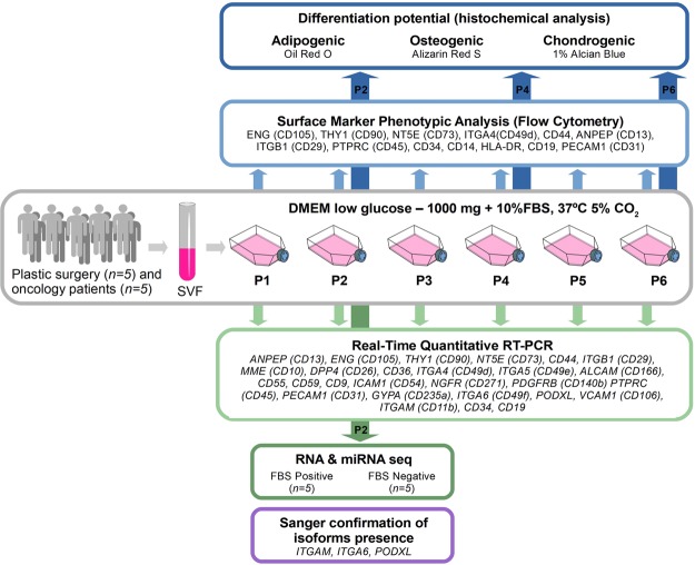
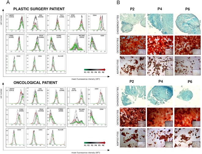
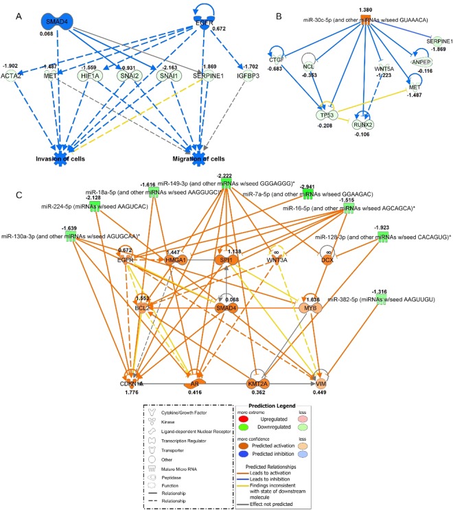
Adipose-derived stem cells (ASCs) have become an important research model in regenerative medicine. However, there are controversies regarding the impact of prolonged cell culture on the ASCs phenotype and their differentiation potential. Hence, we studied 10 clinical ASCs replicates from plastic and oncological surgery patients, in six-passage FBS supplemented cultures. We quantified basic mesenchymal cell surface marker transcripts and the encoded proteins after each passage. In parallel, we investigated the differentiation potential of ASCs into chondrocytes, osteocytes and adipocytes. We further determined the effects of FBS supplementation and subsequent deprivation on the whole transcriptome by comprehensive mRNA and miRNA sequencing. Our results show that ASCs maintain differentiation potential and consistent profile of key mesenchymal markers, with apparent expression of distinct isoforms, in long-term cultures. No significant differences were observed between plastic and oncological surgery cohorts. ASCs in FBS supplemented primary cultures are almost committed to mesenchymal lineages as they express key epithelial-mesenchymal transition genes including early mesenchymal markers. Furthermore, combined mRNA/miRNA expression profiling strongly supports a modulatory role for the miR-30 family in the commitment process to mesenchymal lineages. Finally, we propose improvements to existing qPCR based assays that address alternative isoform expression of mesenchymal markers.


Conflict of interest statement

The authors declare no competing interests.

Figures

Figure 1
Figure 1
Schematic outline of the experimental workflow. ASCs were isolated from human subcutaneous adipose tissue collected from plastic surgery (n = 5) and oncological patients (n = 5). Stem cells obtained from SVF (stromal vascular fraction) were cultured up to the 6th passage. The study consisted of five main phases: (i) ASCs immunophenotype determination using Flow Cytometry Analysis for 13 surface (P1 - P6), (ii) examination of ASCs potential to differentiate to adipocytes, osteocytes and chondrocytes after P2, P4 and P6, (iii) Real-Time Quantitative RT-PCR analysis for 27 ASCs positive and negative “stemness” markers made after each passage (P1–P6), (iv) RNA-seq and miRNA-seq analysis made for 5 replicates after P2 in two experimental conditions: standard (FBS Positive) and serum-deprived (FBS Negative), (v) Sanger sequencing verification of RNA-seq analysis for three transcripts (ITGAM, ITGA6, PODXL) for which significant isoform switching was identified. Further details of the experimental procedure are described in the materials and method section. This figure utilizes modified clipart elements from https://openclipart.org that are covered by Creative Commons Zero 1.0 Public Domain License (http://creativecommons.org/publicdomain/zero/1.0/ and https://openclipart.org/share).
Figure 2
Figure 2
Phenotype identification (A) and assessment of differentiation potential (B) of ASCs originating from plastic and oncological surgery patients. (A) Phenotyping of ASCs standard and supplementary markers using flow cytometry (FCM). FCM plots are shown for two representative patients: plastic and oncological surgery. The X axis represents mean fluorescence intensity (MFI), Y axis - cell number. (B) Representative example of histochemical analysis of ASCs differentiation into chondrocytes, osteocytes and adipocytes from plastic surgery and oncological patients. Differentiation assays were performed after 2nd (P2), 4th (P4), and 6th (P6) passage, oil red O, alizarin red S and 1% alcian blue staining were used to confirm adipocytes, osteocytes and chondrocytes differentiation respectively.
Figure 3
Figure 3
The phenotypic analysis of cultured ASCs. The results are based on 10 biological replicates, including 5 plastic surgery and 5 oncological surgery patients. Flow cytometry assessment of both positive (A) and negative (B) ASCs markers showed statistically significant changes in the expression of the following positive markers: ANPEP (CD13), CD44, NT5E (CD73) and ENG (CD105); and two negative surface markers of ASC – PECAM1 (CD31) and CD34 (all marked with*). The X axis represents the passage number (from P1 to P6), whereas the Y axis displays the median fluorescence intensity (MFI) of cells expressing particular surface marker. The Wilcoxon signed-rank test was used to compare the dynamics of changes between passages. Statistical significance was accepted when P was ≤ 0.01. Center lines denote the median, box limits indicate the 25th and 75th percentiles; whiskers represent the maximum and minimum of the acquired values.
Figure 4
Figure 4
Quantitative PCR gene expression heat maps of primary positive (#) and primary negative (##), secondary positive and negative, non-classical positive and negative ASCs markers. Customary CD nomenclature is supplemented with the official gene names in parentheses. Color intensity depicts transcript quantity, i.e. median expression of target to geometric mean of HPRT1, RPLP0 and RPL13A reference genes expression(T/R values) from white (“0”) to dark blue (“2”). Median value from 5 biological replicates in each group is shown for every passage (P1-P6) for two examined groups of patients from plastic (5 individuals) and oncological surgery (5 individuals).
Figure 5
Figure 5
Networks generated in Ingenuity Pathway Analysis (IPA) for ASCs cultured in standard FBS-supplemented and FBS-deprived conditions for five biological replicates (plastic surgery patients, Supplementary Table S1). The values next to the gene names refer to log2 fold change of gene expression calculated in the cufflinks package. The shapes and colors of molecules and type of interactions are defined in the graphical legends. (A) Top Regulator Effects network based on differentially expressed genes in both experimental conditions with highest consistency score of 3.024. The middle row consists of molecules that are connected to upstream regulators in the top row and downstream functions in the bottom row. (B) miRNA-mRNA negative correlation network with upregulated mir-30 family and anti-correlated mRNA targets generated in microRNA Target Filter Tool in IPA. (C) miRNA-mRNA negative correlation network with downregulated 8 miRNAs and their upregulated direct targets. miRNAs indicated with a star refer to miRNA families that share common seed sequence with those significantly changed in our dataset.

References

    1. Cheng K-H, Kuo T-L, Kuo K-K, Hsiao C-C. Human adipose-derived stem cells: Isolation, characterization and current application in regeneration medicine. Genomic Med. Biomarkers, Heal. Sci. 2011;3:53–62. doi: 10.1016/j.gmbhs.2011.08.003. - DOI
    1. Pikuła M, Marek-Trzonkowska N, Wardowska A, Renkielska A, Trzonkowski P. Adipose tissue-derived stem cells in clinical applications. Expert Opin. Biol. Ther. 2013;13:1357–1370. doi: 10.1517/14712598.2013.823153. - DOI - PubMed
    1. Di Battista, J. A. et al. Proliferation and differentiation of human adipose-derived mesenchymal stem cells (ASCs) into osteoblastic lineage are passage dependent. Inflamm. Res. 907–917, 10.1007/s00011-014-0764-y (2014). - PubMed
    1. Li J, Huang H, Xu X. Biological characteristics and karyotiping of a new isolation method for human adipose mesenchymal stem cells in vitro. Tissue Cell. 2017;49:376–382. doi: 10.1016/j.tice.2017.04.005. - DOI - PubMed
    1. Bieback K, et al. Human alternatives to fetal bovine serum for the expansion of mesenchymal stromal cells from bone marrow. Stem Cells. 2009;27:2331–2341. doi: 10.1002/stem.139. - DOI - PubMed

Publication types