. 2018 Jul 27;8(1):11324.
doi: 10.1038/s41598-018-29745-4.

ClpB mutants of Francisella tularensis subspecies holarctica and tularensis are defective for type VI secretion and intracellular replication

Affiliations

ClpB mutants of Francisella tularensis subspecies holarctica and tularensis are defective for type VI secretion and intracellular replication

Athar Alam et al. Sci Rep. .

Abstract

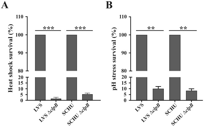
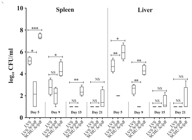
Francisella tularensis, a highly infectious, intracellular bacterium possesses an atypical type VI secretion system (T6SS), which is essential for the virulence of the bacterium. Recent data suggest that the HSP100 family member, ClpB, is involved in T6SS disassembly in the subspecies Francisella novicida. Here, we investigated the role of ClpB for the function of the T6SS and for phenotypic characteristics of the human pathogenic subspecies holarctica and tularensis. The ∆clpB mutants of the human live vaccine strain, LVS, belonging to subspecies holarctica, and the highly virulent SCHU S4 strain, belonging to subspecies tularensis, both showed extreme susceptibility to heat shock and low pH, severely impaired type VI secretion (T6S), and significant, but impaired intracellular replication compared to the wild-type strains. Moreover, they showed essentially intact phagosomal escape. Infection of mice demonstrated that both ΔclpB mutants were highly attenuated, but the SCHU S4 mutant showed more effective replication than the LVS strain. Collectively, our data demonstrate that ClpB performs multiple functions in the F. tularensis subspecies holarctica and tularensis and its function is important for T6S, intracellular replication, and virulence.


Conflict of interest statement

The authors declare no competing interests.

Figures

Figure 1
Figure 1
Survival of indicated F. tularensis strains during heat and pH stress. Overnight grown bacteria were diluted in fresh growth media and subjected to stress at 50 °C for 30 min (A) or pH 4.5 for 1 h (B). Survival of each strain was monitored as viable bacteria and plotted as percentage of survival, setting wild-type strains survival as 100%. There was no significant killing of any wild-type strain during the treatment. Data are the average survival percentage for at least three independent experiments for each condition. Values are mean ± SD. **P < 0.01; *** indicates P < 0.001 vs. the corresponding wild-type strain.
Figure 2
Figure 2
Intracellular growth and LDH release from F. tularensis-infected BMDM and J774A.1 cells. Growth of the LVS or SCHU S4 ΔclpB mutants and the corresponding wild type strains were analyzed by lysis of infected BMDM (A) and J774A.1 (C) cells at 0 h and 18 h and the number of CFU determined. The net growth mean values ± SEM of at least three independent experiments are shown. Supernatants of cultures of BMDM (B) and J774A.1 (D) cells infected with the indicated strains were harvested at 18 h. The activity was expressed as a percentage of the level of uninfected lysed cells (positive lysis control). The mean values ± SEM of triplicate wells from one representative experiment of two are shown. The asterisk indicates that the values of the ∆clpB mutants differed significantly compared to wild type strains (*P < 0.05; **P < 0.01; ***P < 0.001). Unpaired t-test with Welch’s correction was used to compare values.
Figure 3
Figure 3
Co-localization of GFP-expressing strains of F. tularensis with LAMP-1. BMDM cells were infected with the indicated strains expressing GFP for 2 h at an MOI of 30. After 3 and 6 h, specimens were labeled for the late endosomal and lysosomal marker LAMP-1. At least 100 infected cells from multiple cover slips were examined in each experiment. Co-localization was analyzed using a fluorescence microscope (Nikon Eclipse 90i, Nikon, Japan). Results are representative of two experiments and presented as % of co-localization of GFP-expressing bacteria with LAMP-1. The asterisks indicate that the values of the LVS ∆iglC mutant differed significantly compared to LVS at 3 h and 6 h (**P < 0.01); however, the differences between LVS and LVS ∆clpB or SCHU ∆clpB were non-significant (NS P > 0.05). Unpaired t-test with Welch’s correction was used to compare values.
Figure 4
Figure 4
Assessment of the phagosomal membrane integrity of BMDM cells infected with indicated F. tularensis strains by the TEM assay. BMDM cells were infected for 2 h at an MOI of 1,000, washed, further incubated for 3 and 6 h, and then processed for TEM. To examine the membrane integrity of phagosome, at least 100 bacteria from different sections were analyzed for each time point and categorized as follows: Intact, >90% of the phagosomal membrane intact; Slightly damaged, 50–90% of the membrane intact; Highly damaged, 10–50% of the membrane intact; No membrane, <10% of the membrane intact.
Figure 5
Figure 5
Representative examples of secretion of β-lactamase-tagged IglE protein in J774A.1 macrophages. Macrophages were infected either with LVS, LVS ∆clpB, SCHU ∆ggt or SCHU ∆clpB expressing IglE-TEM fusions for 18 h and the fluorescence was determined. TEM β-lactamase activity was identified by the blue fluorescence emitted by the cleaved CCF2 product, whereas uncleaved CCF2 substrate emitted a green fluorescence. The experiments were repeated at least 4 times using duplicate samples. Representative images of each of 4 samples are shown.
Figure 6
Figure 6
Replication of F. tularensis strains in liver and spleen following infection of mice. After intradermal inoculation with 1 × 104 CFU of the indicated strain, mice were sacrificed on day 5, 9, 15 and 21, and bacterial burdens in spleen and liver were determined. The mean ± SEM for six mice per group and time point are shown. A significant difference in the bacterial numbers of mutant strains vs. LVS is indicated as follows: *P < 0.05; **P < 0.01; ***P < 0.001; NS P > 0.05.

References

    1. Sjöstedt A. Tularemia: history, epidemiology, pathogen physiology, and clinical manifestations. Ann N Y Acad Sci. 2007;1105:1–29. doi: 10.1196/annals.1409.009. - DOI - PubMed
    1. Oyston PC, Sjöstedt A, Titball RW. Tularaemia: bioterrorism defence renews interest in Francisella tularensis. Nat Rev Microbiol. 2004;2:967–978. doi: 10.1038/nrmicro1045. - DOI - PubMed
    1. Bröms JE, Sjöstedt A, Lavander M. The role of the Francisella tularensis pathogenicity island in Type VI secretion, intracellular survival, and modulation of host cell signaling. Front Microbiol. 2010;1:136. doi: 10.3389/fmicb.2010.00136. - DOI - PMC - PubMed
    1. Celli J, Zahrt TC. Mechanisms of Francisella tularensis intracellular pathogenesis. Cold Spring Harb Perspect Med. 2013;3:a010314. doi: 10.1101/cshperspect.a010314. - DOI - PMC - PubMed
    1. Hachani A, Wood TE, Filloux A. Type VI secretion and anti-host effectors. Curr Opin Microbiol. 2016;29:81–93. doi: 10.1016/j.mib.2015.11.006. - DOI - PubMed

Publication types

LinkOut - more resources