Review
. 2018 Sep;19(9):535-551.
doi: 10.1038/s41583-018-0039-7.

Brain circuit dysfunction in post-traumatic stress disorder: from mouse to man

Affiliations
Review

Brain circuit dysfunction in post-traumatic stress disorder: from mouse to man

Robert J Fenster et al. Nat Rev Neurosci. 2018 Sep.

Abstract

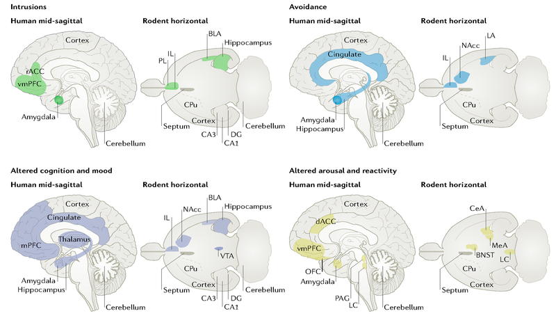
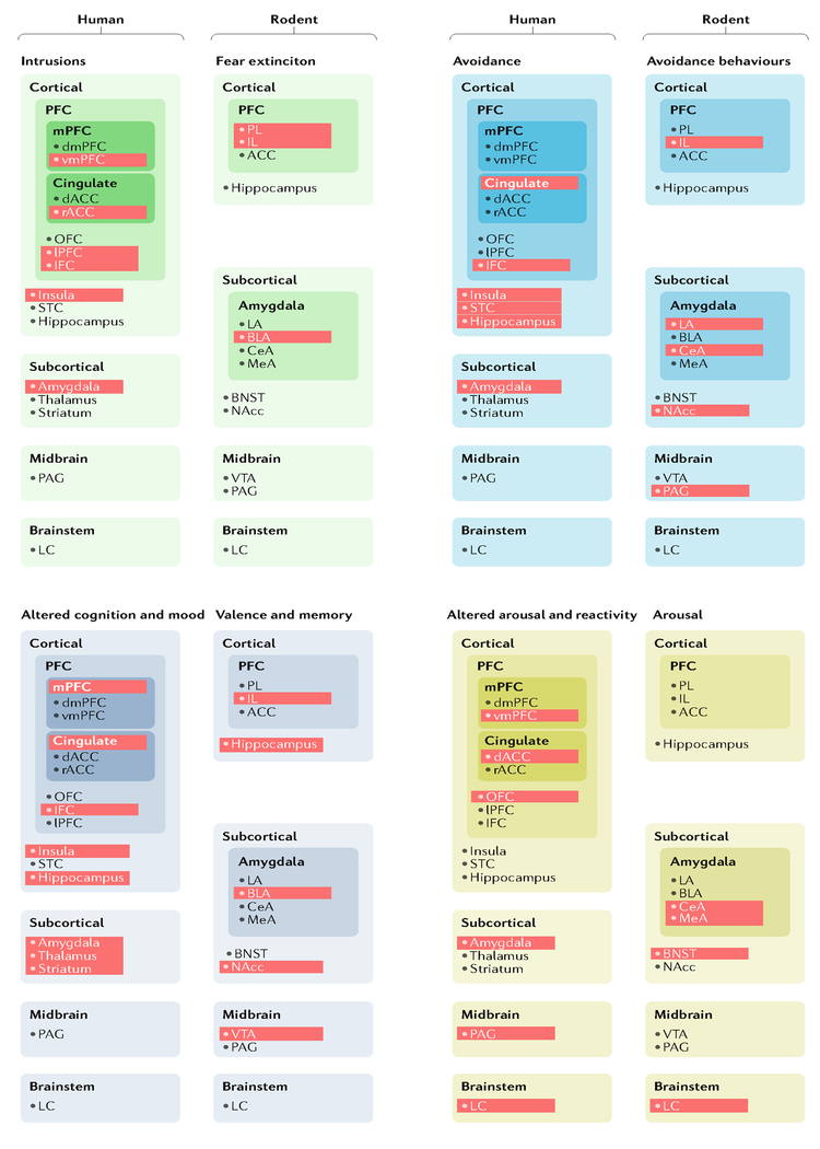
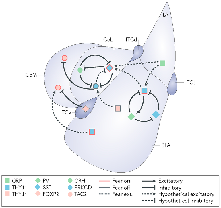
Post-traumatic stress disorder (PTSD) is a prevalent, debilitating and sometimes deadly consequence of exposure to severe psychological trauma. Although effective treatments exist for some individuals, they are limited. New approaches to intervention, treatment and prevention are therefore much needed. In the past few years, the field has rapidly developed a greater understanding of the dysfunctional brain circuits underlying PTSD, a shift in understanding that has been made possible by technological revolutions that have allowed the observation and perturbation of the macrocircuits and microcircuits thought to underlie PTSD-related symptoms. These advances have allowed us to gain a more translational knowledge of PTSD, have provided further insights into the mechanisms of risk and resilience and offer promising avenues for therapeutic discovery.


Conflict of interest statement

Competing interests

K.J.R. is on the scientific advisory boards for Resilience Therapeutics, the Sheppard Pratt-Lieber Research Institute, the Laureate Institute for Brain Research, the Army Study to Assess Risk and Resilience in Servicemembers (STARRS) project, the University of California-San Diego VA Center of Excellence for Stress and Mental Health (CESAMH) and the Anxiety and Depression Association of America; provides fee-for-service consultation for Biogen and Resilience Therapeutics; and holds patents for the use of d-cycloserine and psychotherapy, targeting the pituitary adenylate cyclase-activating polypeptide (PACAP) type 1 receptor for extinction, targeting tachykinin 2 for prevention of fear and targeting angiotensin to improve extinction of fear. R.J.F., L.A.M.L. and J.S. declare no competing interests.

Figures

Fig. 1|
Fig. 1|. An expanded neurocircuitry of post-traumatic stress disorder.
Recent work suggests that an expanded brain network is implicated in post-traumatic stress disorder (PTSD) symptoms. This figure highlights brain regions that have been implicated in either human imaging studies of PTSD (mid-sagittal section) or in rodent models of related behaviours (horizontal section). Each quadrant illustrates brain regions that have evidence linking them to symptoms in the labelled cluster. Rodent behaviours were chosen that map aspects of the corresponding symptom cluster: fear extinction (intrusions); active and passive avoidance (avoidance); spatial memory, emotional valence and anhedonia (altered cognition and mood); and aggression and arousal (altered reactivity and arousal). BLA, basolateral amygdala; BNST, bed nucleus of the stria terminals; CeA, central amygdala; CPu, caudate and putamen; dACC, dorsal anterior cingulate cortex; DG, dentate gyrus; IL, infralimbic cortex; LA, lateral amygdala; LC, locus coeruleus; MeA, medial amygdala; mPFC, medial prefrontal cortex; NAcc, nucleus accumbens; OFC, orbitofrontal cortex; PAG, periaqueductal grey; PL, prelimbic cortex; rACC, rostral anterior cingulate cortex; vmPFC, ventromedial prefrontal cortex; VTA, ventral tegmental area.
Fig. 2|
Fig. 2|. A map of post-traumatic stress disorder neurocircuits in humans and rodent models.
The schematic illustrates the brain regions currently implicated in each of the four major post-traumatic stress disorder (PTSD) symptom clusters by experiments involving human neuroimaging or rodent models. Red shading indicates areas for which there is evidence linking that particular brain region to the symptom cluster or related rodent behaviour. ACC, anterior cingulate cortex; BLA, basolateral amygdala; BNST, bed nucleus of the stria terminals; CeA, central amygdala; dACC, dorsal anterior cingulate cortex; dmPFC, dorsomedial prefrontal cortex; IFC, inferior frontal cortex; IL, infralimbic cortex; LA, lateral amygdala; LC, locus coeruleus; LPFC, lateral prefrontal cortex; MeA, medial amygdala; mPFC, medial prefrontal cortex; NAcc, nucleus accumbens; OFC, orbitofrontal cortex; PAG, periaqueductal grey; PFC, prefrontal cortex; PL, prelimbic cortex; rACC, rostral anterior cingulate cortex; STC, superior temporal cortex; vmPFC, ventromedial prefrontal cortex; VTA, ventral tegmental area.
Fig. 3|
Fig. 3|. Amygdala microcircuits implicated in fear conditioning.
The schematic offers a simplified representation of the mouse amygdala microcircuits that are currently implicated in fear conditioning. During fear conditioning, output neurons from the central amygdala increase their responsiveness to a conditioned stimulus. This increased responsiveness is likely to occur through the mutual interaction of two parallel ‘fear on’ and ‘fear off’ pathways that project to these neurons from the basolateral amygdala (BLA) and lateral amygdala (LA). There is an additional pathway for the establishment of competing fear extinction memories that is likely to originate from neurons in the infralimbic cortex that activate intercalated cells in the amygdala (‘fear ext.’ neurons). Although our understanding of these pathways remains incomplete, several cell types have been demonstrated to modify the expression of fear behaviours. Excitatory connections are indicated with arrows. Inhibitory connections are indicated with blunt arrows. Solid lines designate proven connections, whereas dashed lines illustrate hypothetical connections. CeL, lateral division of the central amygdala; CeM, medial division of the central amygdala; CRH, corticotropin-releasing hormone; FOXP2, forkhead box protein P2; GRP, gastrin-releasing peptide; ITCd, ITCv and ITCl, intercalated cell masses dorsal, ventral and lateral, respectively; PRKCD, protein kinase C delta type; PV, parvalbumin; SST, somatostatin; TAC2, protachykinin 1 (also known as TAC1); THY1, Thy-1 membrane glycoprotein.

References

    1. Morris DJ The Evil Hours: A Biography of Post-Traumatic Stress Disorder. (Houghton Mifflin Harcourt, 2015).
    1. van der Kolk B Interview: what is PTSD really? Surprises, twists of history, and the politics of diagnosis and treatment. Interview by Lisa M Najavits. J. Clin. Psychol 69, 516–522 (2013). - PubMed
    1. American Psychiatric Association. Diagnostic and statistical manual of mental disorders (DSM-3). (APA Publishing, 1980).
    1. Logue MW et al. The Psychiatric Genomics Consortium Posttraumatic Stress Disorder Workgroup: posttraumatic stress disorder enters the age of large-scale genomic collaboration. Neuropsychopharmacology 40, 2287–2297 (2015). - PMC - PubMed
    1. Insel T et al. Research domain criteria (RDoC): toward a new classification framework for research on mental disorders. Am. J. Psychiatry 167, 748–751 (2010). - PubMed

Publication types