

Upstream stimulating factor 1 suppresses autophagy and hepatic lipid droplet catabolism by activating mTOR
- PMID: 30054905
- PMCID: PMC6175420
- DOI: 10.1002/1873-3468.13203
Upstream stimulating factor 1 suppresses autophagy and hepatic lipid droplet catabolism by activating mTOR
Abstract
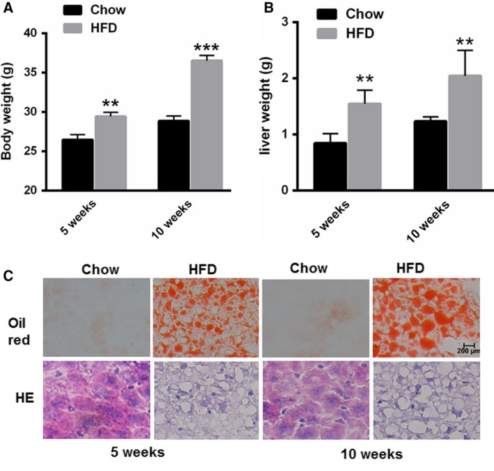
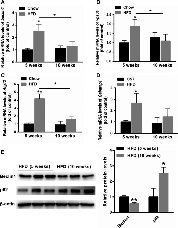
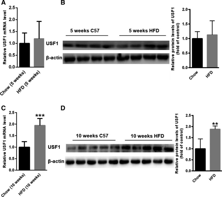
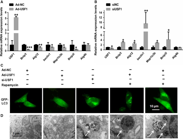
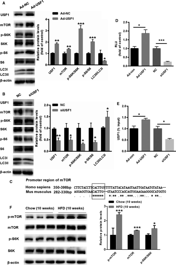
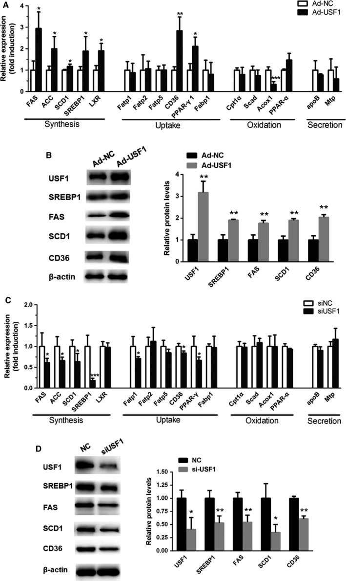
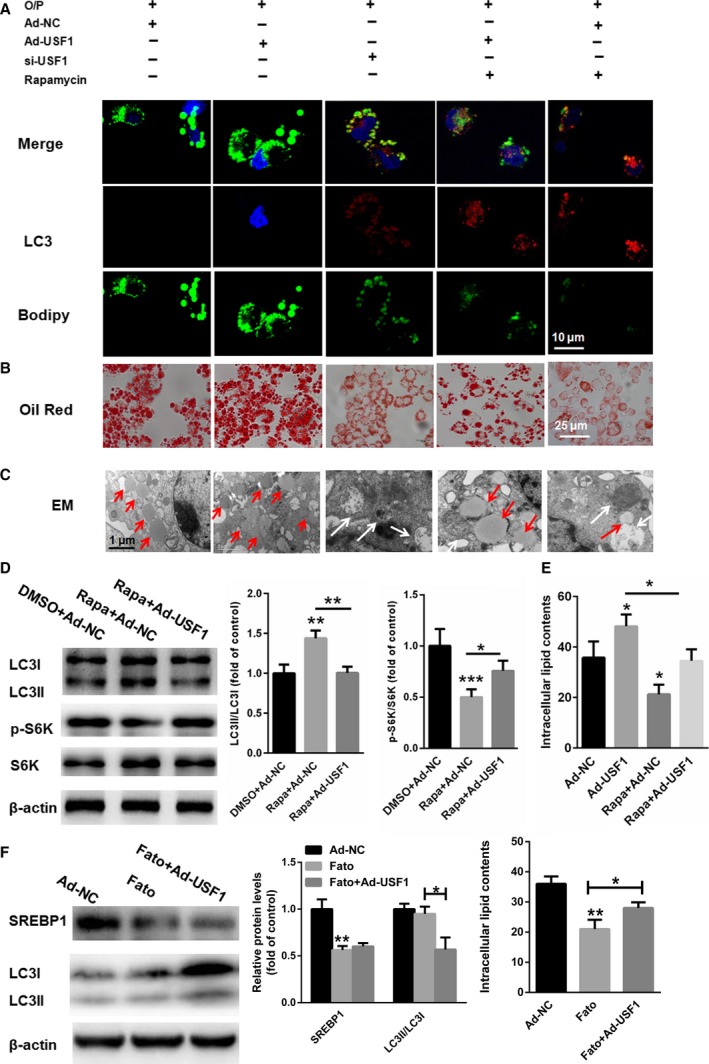
Previous studies indicate that the transcription factor upstream stimulating factor 1 (USF1) is involved in the regulation of lipid and glucose metabolism. However, the role of USF1 in lipid-induced autophagy remains unknown. Interestingly, we found that USF1 overexpression suppresses autophagy-related gene expression in HepG2 cells. Further assays confirmed that USF1 could transcriptionally activate mTOR expression, thereby suppressing rapamycin-induced autophagy in HepG2 cells. Moreover, pharmacological activation of autophagy with rapamycin decreases the numbers and sizes of lipid droplets (LDs) in HepG2 cells exposed to an oleate/palmitate mixture. Of note, USF1 upregulation decreases colocalization of LDs and autophagosomes. In conclusion, our data provide evidence that USF1 contributes to abnormal lipid accumulation in the liver by suppressing autophagy via regulation of mTOR transcription.
Keywords: USF1; autophagy; lipid droplet catabolism; mTOR.
© 2018 The Authors. FEBS Letters published by John Wiley & Sons Ltd on behalf of Federation of European Biochemical Societies.
Figures
Similar articles
-
Resveratrol Ameliorates Lipid Droplet Accumulation in Liver Through a SIRT1/ ATF6-Dependent Mechanism.Cell Physiol Biochem. 2018;51(5):2397-2420. doi: 10.1159/000495898. Epub 2018 Dec 11. Cell Physiol Biochem. 2018. PMID: 30537742
-
Role of USF1 in activating CYBA transcription and influencing NADPH-ROS-mediated oxidative stress and lipid accumulation in non-alcoholic fatty liver disease.Biochim Biophys Acta Mol Cell Biol Lipids. 2025 Mar;1870(2):159581. doi: 10.1016/j.bbalip.2024.159581. Epub 2024 Nov 20. Biochim Biophys Acta Mol Cell Biol Lipids. 2025. PMID: 39577491
-
Cordycepin alleviates hepatic lipid accumulation by inducing protective autophagy via PKA/mTOR pathway.Biochem Biophys Res Commun. 2019 Aug 27;516(3):632-638. doi: 10.1016/j.bbrc.2019.06.108. Epub 2019 Jun 23. Biochem Biophys Res Commun. 2019. PMID: 31242974
-
Ghrelin Attenuated Lipotoxicity via Autophagy Induction and Nuclear Factor-κB Inhibition.Cell Physiol Biochem. 2015;37(2):563-76. doi: 10.1159/000430377. Epub 2015 Sep 1. Cell Physiol Biochem. 2015. PMID: 26329041
-
Lipid Droplet Formation and Lipophagy in Fatty Liver Disease.Semin Liver Dis. 2019 Jul;39(3):283-290. doi: 10.1055/s-0039-1685524. Epub 2019 Apr 30. Semin Liver Dis. 2019. PMID: 31041790 Free PMC article. Review.
Cited by
-
Upregulation of Superenhancer-Driven LncRNA FASRL by USF1 Promotes De Novo Fatty Acid Biosynthesis to Exacerbate Hepatocellular Carcinoma.Adv Sci (Weinh). 2022 Oct 28;10(1):e2204711. doi: 10.1002/advs.202204711. Online ahead of print. Adv Sci (Weinh). 2022. PMID: 36307901 Free PMC article.
-
USF1 modulates transcription and cellular functions by regulating multiple transcription factors in Huh7 cells.Oncol Lett. 2023 Oct 30;26(6):532. doi: 10.3892/ol.2023.14119. eCollection 2023 Dec. Oncol Lett. 2023. PMID: 38020298 Free PMC article.
-
Nasal Septum Deviation as the Consequence of BMP-Controlled Changes to Cartilage Properties.Front Cell Dev Biol. 2021 Jun 24;9:696545. doi: 10.3389/fcell.2021.696545. eCollection 2021. Front Cell Dev Biol. 2021. PMID: 34249945 Free PMC article.
-
The ménage à trois of autophagy, lipid droplets and liver disease.Autophagy. 2022 Jan;18(1):50-72. doi: 10.1080/15548627.2021.1895658. Epub 2021 Apr 2. Autophagy. 2022. PMID: 33794741 Free PMC article. Review.
-
An in silico approach to identify potential downstream targets of miR-153 involved in Alzheimer's disease.Front Genet. 2024 Jan 16;15:1271404. doi: 10.3389/fgene.2024.1271404. eCollection 2024. Front Genet. 2024. PMID: 38299037 Free PMC article.
References
-
- Papackova Z, Dankova H, Palenickova E, Kazdova L and Cahova M (2012) Effect of short‐ and long‐term high‐fat feeding on autophagy flux and lysosomal activity in rat liver. Physiol Res 61 (Suppl 2), S67–S76. - PubMed
Publication types
MeSH terms
Substances
LinkOut - more resources
Full Text Sources
Other Literature Sources
Medical
Miscellaneous
