. 2018 Aug 23;174(5):1264-1276.e15.
doi: 10.1016/j.cell.2018.06.036. Epub 2018 Jul 26.

Progenitor Hyperpolarization Regulates the Sequential Generation of Neuronal Subtypes in the Developing Neocortex

Affiliations

Progenitor Hyperpolarization Regulates the Sequential Generation of Neuronal Subtypes in the Developing Neocortex

Ilaria Vitali et al. Cell. .

Abstract

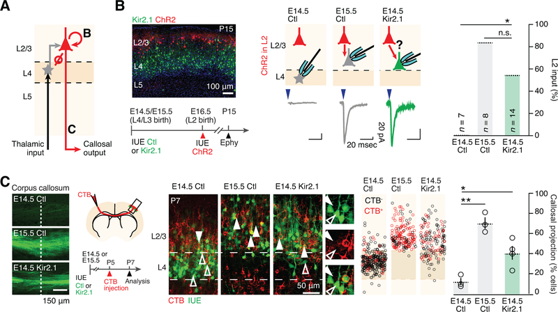
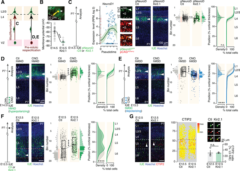
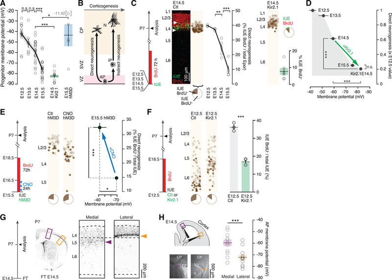
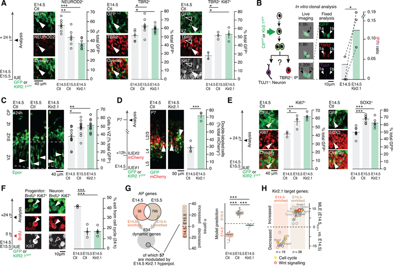
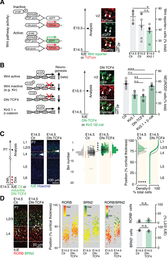
During corticogenesis, ventricular zone progenitors sequentially generate distinct subtypes of neurons, accounting for the diversity of neocortical cells and the circuits they form. While activity-dependent processes are critical for the differentiation and circuit assembly of postmitotic neurons, how bioelectrical processes affect nonexcitable cells, such as progenitors, remains largely unknown. Here, we reveal that, in the developing mouse neocortex, ventricular zone progenitors become more hyperpolarized as they generate successive subtypes of neurons. Experimental in vivo hyperpolarization shifted the transcriptional programs and division modes of these progenitors to a later developmental status, with precocious generation of intermediate progenitors and a forward shift in the laminar, molecular, morphological, and circuit features of their neuronal progeny. These effects occurred through inhibition of the Wnt-beta-catenin signaling pathway by hyperpolarization. Thus, during corticogenesis, bioelectric membrane properties are permissive for specific molecular pathways to coordinate the temporal progression of progenitor developmental programs and thus neocortical neuron diversity.

Keywords: cortical development; membrane potential; neuronal diversity; progenitors.


Conflict of interest statement

DECLARATION OF INTERESTS

No competing interests to declare.

Figures

Figure 1
Figure 1
Kir2.1 electroporation at E14.5 leads to premature presence of L2/3-type neurons. (A) The radial location of neurons born in the Kir2.1-electroporation condition shifts superficially towards that of normally later born neurons. (B) Possible scenarios explaining the radial shift. (C) Left: RORB and BRN2 are expressed along opposing radial gradients in L4 and L2/3. Middle and right: Molecular expression is congruent with laminar location in single neurons in Kir2.1-electroporation condition. (D) Fluorescently-labeled neurons collected at P3 for transcriptomic analysis. (E) Neurons repress L4 neuron-type and induce L2/3 neuron-type genes in the Kir2.1-electroporation condition. (F) Left: unbiased SVM classification reveals a shift of the transcriptional identity of E14.5-born neurons in the Kir2.1-electroporation. Middle: Top 20 genes used to build the model. Right: Expression of selected genes; in situ hybridizations from Allen Brain Atlas. Data are represented as mean ± SEM, except for scatter plots represented as means ± SD. (A), two-way ANOVA for bin distribution analysis (significantly different bins indicated in darker shades); Student’s t test for scatter plot (comparing average cell position). Individual biological replicates are distinguished by color and aligned from left to right. (C), Student’s t test. (F), one-way ANOVA. *: P<0.05; **: P<10−2; ***: P<10−3; ****: P<10−4. Ctl: Control; Epor: electroporated cells; VZ: ventricular zone. See also Figure S1.
Figure 2
Figure 2
Kir2.1 in utero electroporation leads to a forward shift in neuronal morphology. (A) In the Kir2.1-electroporation condition, neurons display a non-polarized dendritic arbor, as E15.5-born L2/3-type neurons do. Right: cumulative radial distribution of primary dendrites for each analyzed cell. (B) E14.5-born neurons mostly display a stellate morphology whereas Kir2.1-electroporated neurons often display an apical dendrite, as do E15.5-born L2/3-type neurons (red arrowheads). Horizontal bars in (A) indicate median values. Data in (B) are represented as means ± SEM. (A), Kruskal-Wallis test; (B), one-way ANOVA. *: P<0.05; **: P<10−2; ***: P<10−3.
Figure 3
Figure 3
Kir2.1 in utero electroporation leads to a L4-to-L2/3 shift in input-output circuit properties. (A) Schematic representation of input-output connectivity in S1. Letters refer to panels. (B) In the Kir2.1-electroporation condition, neurons acquire L2/3 neuron-type intracortical input. (C) Kir2.1-electroporated neurons acquire L2/3 neuron-type intracortical output. Left: axons of electroporated neurons are visible in the corpus callosum. Dashed line corresponds to midline. Data are represented as mean ± SEM. (B) Fisher’s exact test; (C) one-way ANOVA. *: P<0.05; **: P<10−2. IUE: in utero electroporation; ChR2: channelrhodopsin; CTB: cholera toxin B. See also Figure S2.
Figure 4
Figure 4
Apical progenitors precociously generate next-born neuronal subtypes following Kir2.1-induced hyperpolarization. (A) Possible scenarios explaining the laminar shift. Reported from Figure 1B. Letters refer to panels. (B) Kir2.1 overexpression lowers the Vm of E14.5 APs. Inset: Fluorescent dye-filled patched AP (arrowhead). (C) Left: Gene expression dynamics for NeuroD1 (from Telley et al., 2016, see http://genebrowser.unige.ch/science2016). Right: Postmitotic expression of Kir2.1 does not affect the radial positioning of E14.5-born neurons. (D) Premitotical hyperpolarization by early CNO pulse-injection in hM4D-expressing APs shifts the laminar location of E14.5-born neurons towards that of normally later-born neurons. (E) Premitotical depolarization by early CNO pulse-injection in hM3D-expressing APs shifts the radial location of E15.5-born neurons towards that of normally earlier-born neurons. (F) The radial distribution of E12.5-born neurons following Kir2.1 hyperpolarization is shifted towards that of later-born neurons. This shift is restricted to deep-layer neurons, a time at which the Kir2.1-expression plasmid is most concentrated. (G) Molecular expression of the L5B marker CTIP2 is congruent with radial shift in location following Kir2.1 hyperpolarization. The percentage of cells expressing CTIP2 in L5B is unchanged in control and Kir conditions. Data are represented as mean ± SEM, except scatter plots represented as means ± SD. (B), one-way ANOVA; (C-F), two-way ANOVA for bin distribution analysis (significantly different bins indicated in darker shades); Student’s t test for scatter plot (comparing average cell position). Individual biological replicates are distinguished by color and aligned from left to right. (G), Student’s t test. ***: P<10−3; ****: P<10−4. SVZ: subventricular zone. See also Figure S3.
Figure 5
Figure 5
Apical progenitor hyperpolarization regulates the progression from direct to indirect neurogenic divisions. (A) APs hyperpolarize in the course of corticogenesis. Kir2.1 overexpression and CNO activation of hM3D-expressing APs induce hyperpolarization and depolarization respectively. E14.5 Kir2.1 and E15.5 hM3D data reported from Figures 4B and S3A, respectively. (B) Schematic representation of the shift from direct to indirect neurogenesis during corticogenesis. Note that indirect neurogenesis is also present at lower levels at early stages. (C) Direct neurogenesis decreases during corticogenesis. Directly-born neurons are Epor+BrdU. Kir2.1-hyperpolarization decreases direct neurogenesis in E14.5 APs. Note that while this approach identifies directly-born neurons as GFP+BrdU cells, GFP+BrdU+ cells cannot be assumed to be born indirectly, since they may be born directly from another round of progenitor divisions (Telley et al., 2016). (D) Summary of the findings: Kir2.1 overexpression hyperpolarizes APs and leads to a decrease in direct neurogenesis. (E) Depolarization of hM3D-expressing APs by CNO pulse-injection increases direct neurogenesis. (F) Kir2.1-hyperpolarization decreases direct neurogenesis in E12.5 APs. (G) At E14.5, medial cortical areas produce L5 neurons, while lateral areas produce L4 neurons. P7 brains following FT injection at E14.5, which labels neurons directly produced by APs at that time. (H) Laterally-located APs are more hyperpolarized than medial ones at E14.5, corresponding to the type of neuron being produced. Arrowheads point to the tip of the recording pipette. Data are represented as mean ± SEM. (A), (C), (D) and (F): one-way ANOVA; (E) and (H): Student’s t-test. *: P<0.05; **: P<10−2; ***: P<10−3. CP: cortical plate; FT: FlashTag; V: ventricle; AP: apical progenitor. See also Figure S4.
Figure 6
Figure 6
Hyperpolarization leads to a forward shift in apical progenitor cycling behavior and transcriptional identity. (A) Kir2.1-hyperpolarization of APs decreases neuronal production (NEUROD2+) and increases IP numbers (TBR2+) in vivo. TBR2/Ki67 double labeling is used to highlight newborn IPs in the VZ. (B) In vitro single cell clonal analysis shows increased IP production relative to neurons in the Kir2.1-hyperpolarization condition. Dotted lines connect biological replicates. (C) The proportion of cells that remain in the VZ after E14.5 Kir2.1-hyperpolarization is increased towards E15.5 levels. (D) In contrast to control cells, E14.5 Kir2.1-hyperpolarized cells remain susceptible to targeting by a second IUE 12 hours after the first IUE, suggesting a prolonged pre-mitotic period (white arrowheads show double labeled neurons). (E) E14.5 Kir2.1 hyperpolarization increases the proportion of progenitor cells (Ki67+ and SOX2+). (F) Kir2.1-hyperpolarized APs show decreased cell cycle exit as demonstrated by double labeling for BrdU and Ki67 at 24h. Cells that remain in the cell cycle are BrdU+Ki67+ while neurons are BrdU+Ki67. (G) Left: E14.5 Kir2.1-hyperpolarized APs repress E14.5-type and induce E15.5-type genes 12h after IUE. Right: unbiased SVM classification reveals that the transcriptional identity of E14.5 Kir2.1-hyperpolarized APs is shifted towards E15.5 APs. (H) Differentially-enriched genes include several cell cycle- and Wnt signaling-related transcripts. Data are represented as mean ± SEM. (A), (C), (E-G): one-way ANOVA; (B), (D): Student’s test. IP: intermediate progenitor; N: neuron; IZ: intermediate zone. *: P<0.05; **: P<10−2; ***: P<10−3. See also Figures S5–7.
Figure 7
Figure 7
Membrane hyperpolarization represses Wnt signaling and drives the developmental progression in daughter neuron identity. (A) AP hyperpolarization decreases canonical Wnt pathway activity. Arrowheads show example of 2 cells with active Wnt signaling as revealed by IUE of a Wnt reporter construct. (B) Blocking canonical Wnt signaling by overexpressing the dominant negative form of TCF4 replicates the effect of Kir2.1-mediated hyperpolarization on neuronal output. Inducing canonical Wnt signaling by β-catenin overexpression rescues hyperpolarization-mediated reduction in neuronal output. Control and Kir2.1 values have been reported from Figure 6A for comparison. (C) E14.5 AP-targeted repression of Wnt signaling using an inducible DN-TCF4 construct causes a forward shift in their laminar identity. Compare with Fig. 1A. (D) Molecular expression is congruent with laminar location in single neurons following E14.5 AP Wnt signaling repression. Data are represented as mean ± SEM, except scatter plots represented as means ± SD. (A), (B): one-way ANOVA; (C), two-way ANOVA for bin distribution analysis (statistically significantly different bins indicated in darker shades); Student’s t test for scatter plot (comparing average cell position). Individual biological replicates are distinguished by color and aligned from left to right. (D), Student’s t test. *: P<0.05; **: P<10−2; ***: P<10−3; ****: P<10−4. DN-TCF4: dominant negative TCF4; ND2: NEUROD2.

Comment in

References

    1. Arcangeli A, Bianchi L, Becchetti A, Faravelli L, Coronnello M, Mini E, Olivotto M, and Wanke E (1995). A novel inward-rectifying K+ current with a cell-cycle dependence governs the resting potential of mammalian neuroblastoma cells. J. Physiol. (Lond.) 489 (Pt 2), 455. - PMC - PubMed
    1. Arlotta P, Molyneaux BJ, Chen J, Inoue J, Kominami R, Macklis JD (2005). Neuronal subtype-specific genes that control corticospinal motor neuron development in vivo. Neuron 45, 207–21. - PubMed
    1. Armbruster BN, Li X, Pausch MH, Herlitze S, and Roth BL (2007). Evolving the lock to fit the key to create a family of G protein-coupled receptors potently activated by an inert ligand. Proc. Natl. Acad. Sci. U.S.a 104, 5163–5168. - PMC - PubMed
    1. Ataman B, Ashley J, Gorczyca M, Ramachandran P, Fouquet W, Sigrist SJ, and Budnik V (2008). Rapid activity-dependent modifications in synaptic structure and function require bidirectional WNT signaling. Neuron 57, 705–718. - PMC - PubMed
    1. Bauer CK, and Schwarz JR (2001). Physiology of EAG K+ channels. J. Membr. Biol 182, 1–15. - PubMed

Publication types

MeSH terms