. 2018 Aug 9;174(4):870-883.e17.
doi: 10.1016/j.cell.2018.06.029. Epub 2018 Jul 26.

The Mitochondrial Unfolded Protein Response Is Mediated Cell-Non-autonomously by Retromer-Dependent Wnt Signaling

Affiliations

The Mitochondrial Unfolded Protein Response Is Mediated Cell-Non-autonomously by Retromer-Dependent Wnt Signaling

Qian Zhang et al. Cell. .

Abstract

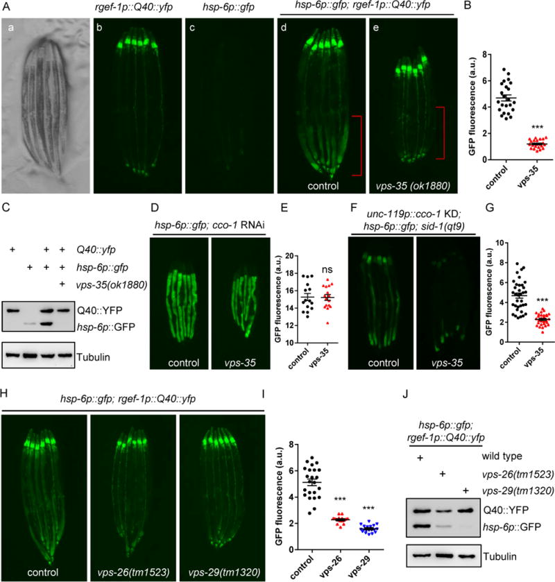
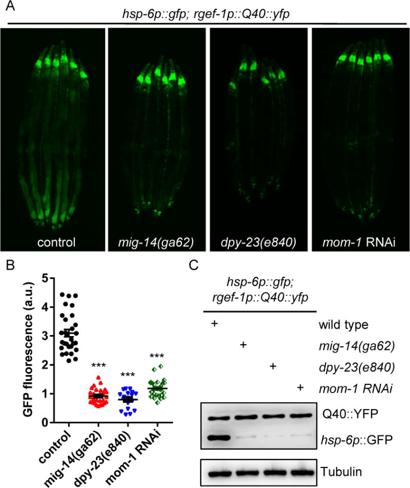
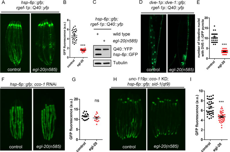
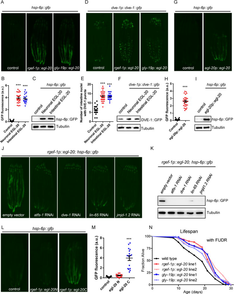
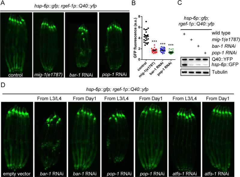
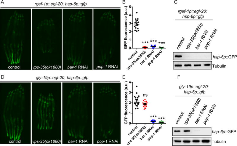
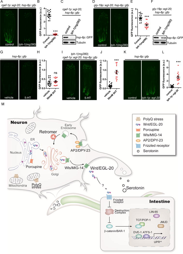
The mitochondrial unfolded protein response (UPRmt) can be triggered in a cell-non-autonomous fashion across multiple tissues in response to mitochondrial dysfunction. The ability to communicate information about the presence of mitochondrial stress enables a global response that can ultimately better protect an organism from local mitochondrial challenges. We find that animals use retromer-dependent Wnt signaling to propagate mitochondrial stress signals from the nervous system to peripheral tissues. Specifically, the polyQ40-triggered activation of mitochondrial stress or reduction of cco-1 (complex IV subunit) in neurons of C. elegans results in the Wnt-dependent induction of cell-non-autonomous UPRmt in peripheral cells. Loss-of-function mutations of retromer complex components that are responsible for recycling the Wnt secretion-factor/MIG-14 prevent Wnt secretion and thereby suppress cell-non-autonomous UPRmt. Neuronal expression of the Wnt ligand/EGL-20 is sufficient to induce cell-non-autonomous UPRmt in a retromer complex-, Wnt signaling-, and serotonin-dependent manner, clearly implicating Wnt signaling as a strong candidate for the "mitokine" signal.

Keywords: EGL-20; UPR(mt); VPS-35; Wnt signaling; mitochondrial unfolded protein response; mitokine; retromer complex.


Conflict of interest statement

Declaration of Interests

A.D. is a cofounder of Proteostasis Therapeutics, Inc. and Mitobridge, Inc. and declares no financial interest related to this work.

Figures

Figure 1
Figure 1. The retromer complex is required for cell-non-autonomous UPRmt induction in animals expressing Q40∷YFP in neurons
(A) Representative photomicrographs demonstrating: a) bright field images of aligned, WT animals; b) Q40∷yfp expression in neurons; c) hsp-6p∷gfp expression in WT animals; d) hsp-6p∷gfp was up-regulated in the intestine in day2 adult animals expressing neuronal Q40∷YFP; e) hsp-6p∷gfp expression was suppressed in vps-35 mutants. The posterior region of the intestine where hsp-6p∷gfp is induced or suppressed is highlighted in (d) and (e). Scale bar: 250μm. (B) Quantification of hsp-6p∷gfp expression of the entire intestine in animals expressing Q40∷YFP in neurons with the presence (d) or absence of the vps-35 mutation (e) as shown in (A). (C) Immunoblots of GFP expression in animals as indicated. (D) Representative photomicrographs of hsp-6p∷gfp animals with the presence or absence of the vps-35 mutation grown on empty vector (EV) or with cco-1 RNAi from hatching. (E) Quantification of hsp-6p∷gfp expression. The genotypes are as in (D). (F) Representative photomicrographs of neuronal cco-1 knockdown; sid-1(qt9); hsp-6p∷gfp animals with the presence or absence of vps-35 mutation. (G) Quantification of hsp-6p∷gfp expression. The genotypes are as in (F). (H) Representative photomicrographs of animals expressing neuronal Q40∷YFP; hsp-6p∷gfp in WT, vps-26, and vps-29 animals. (I) Quantification of hsp-6p∷gfp expression. The genotypes are as in (H). (J) Immunoblots of GFP expression in animals as indicated. YFP can be recognized by the GFP antibody. Anti-tubulin serves as a loading control. *** p < 0.0001, ns denotes p > 0.05 via t-test. Error bars, SEM. n ≥15 worms. See also Figure S1.
Figure 2
Figure 2. Retromer-dependent Wnt secretion is required for cell-non-autonomous UPRmt induction in animals expressing Q40∷YFP in neurons
(A) Representative photomicrographs of animals expressing neuronal Q40∷YFP; hsp-6p∷gfp in WT, mig-14, dpy-23, or mom-1 RNAi animals. (B) Quantifications of the hsp-6p∷gfp expression as shown in (A). *** p < 0.0001 via t test. Error bars, SEM. n ≥20 worms. (C) Immunoblots of GFP expression in animals as indicated. See also Figure S2.
Figure 3
Figure 3. Wnt ligand/EGL-20 is required for cell-non-autonomous UPRmt induction in animals with Q40∷YFP expression in neurons
(A) Representative photomicrographs of D2 adult animals expressing neuronal Q40∷YFP; hsp-6p∷gfp in WT or egl-20 animals. (B) Quantification of hsp-6p∷gfp expression. The genotypes are as in (A). (C) Immunoblots of GFP expression. The genotypes are as in (A). (D) Representative photomicrographs of D2 adult animals expressing neuronal Q40∷YFP; DVE-1∷GFP in WT and egl-20 animals. Arrows highlight the DVE-1∷GFP signal localized in the intestinal nuclei. (E) Quantification of the number of intestinal nuclei with DVE-1 puncta. The genotypes are as in (D). (F) Representative photomicrographs of D1 animals expressing hsp-6p∷gfp in WT or egl-20 animals grown on EV or cco-1 RNAi from hatching. (G) Quantification of hsp-6p∷gfp expression. The genotypes are as in (F). (H) Representative photomicrographs of hsp-6p∷gfp expression in neuronal specific cco-1 RNAi animals in WT or egl-20 background. (I) Quantification of hsp-6p∷gfp expression. The genotypes are as in (H). *** p< 0.0001, ns denotes p > 0.05 via t-test. Error bars, SEM. n ≥ 15 worms. See also Figure S3.
Figure 4
Figure 4. Expression of Wnt ligand/EGL-20 is sufficient to induce UPRmt in both cell-autonomous and cell-non-autonomous manners
(A) Representative photomicrographs of hsp-6p∷gfp reporter animals in WT, rgef-1p∷egl-20 (neuronal), and gly-19p∷egl-20(intestinal) background. (B) Quantification of hsp-6p∷gfp expression. The genotypes are as in (A). (C) Immunoblot of hsp-6p∷gfp expression. The genotypes are as in (A). (D) Representative photomicrographs of DVE-1∷GFP reporter animals in WT, rgef-1p∷egl-20, and gly-19p∷egl-20 background. (E) Quantification of the number of intestinal nuclei with DVE-1 puncta. The genotypes are as in (D). (F) Immunoblot of DVE-1∷GFP expression. The genotypes are as in (D). (G) Representative photomicrographs of hsp-6p∷gfp reporter animals in WT or egl-20p∷egl-20∷mCherry background. (H) Quantification of hsp-6p∷gfp expression. The genotypes are as in (G). (I) Immunoblot of hsp-6p∷gfp expression. The genotypes are as in (G). (J) Representative photomicrographs of rgef-1p∷egl-20; hsp6p∷gfp reporter animals grown on EV, atfs-1, dve-1, lin-65, or jmjd-1.2 RNAi from hatching. (K) Immunoblot of hsp-6p∷gfp expression. The genotypes are as in (J). (L) Representative photomicrographs of hsp-6p∷gfp reporter animals in WT, rgef-1p∷egl-20N(1-720)∷mCherry, or rgef-1p∷egl-20C(721-1182)∷mCherry background. (M) Quantification of hsp-6p∷gfp expression. The genotypes are as in (L). (N) Overexpression of egl-20 in neurons or in the intestine extends C. elegans lifespan. Lifespan analysis of two independent transgenic lines of rgef-1p∷egl-20 or gly-19p∷egl-20 expressing animals compared to WT animals. See also Table S1. *** p< 0.0001 via t-test. Error bars, SEM. n ≥ 20 worms. See also Figure S4.
Figure 5
Figure 5. The canonical Wnt/β-catenin signaling pathway is required for cell-non-autonomous UPRmt induction in animals expressing Q40∷YFP in neurons
(A) Representative photomicrographs of neuronal Q40∷YFP; hsp-6p∷gfp expressing animals in WT, mig-1, bar-1 RNAi, or pop-1 RNAi background. (B) Quantification of hsp-6p∷gfp expression. The genotypes are as in (A). *** p< 0.0001 via t-test. Error bars, SEM. n ≥ 20 worms. (C) Immunoblots of GFP expression. The genotypes are as in (A). (D) Representative photomicrographs of neuronal Q40∷YFP; hsp-6p∷gfp expressing animals transferred to RNAi plates either from L3/L4 stages or D1 adulthood. See also Figure S5.
Figure 6
Figure 6. The induction of cell-non-autonomous UPRmt signaling upon neuronal Wnt/EGL-20 expression is dependent on the retromer complex and canonical Wnt signaling
(A) Representative photomicrographs of rgef-1p∷egl-20; hsp-6p∷gfp reporter animals in WT, vps-35, bar-1 RNAi, or pop-1 RNAi background. (B) Quantification of hsp-6p∷gfp expression. The genotypes are as in (A). (C) Immunoblot of GFP expression. The genotypes are as in (A). (D) Representative photomicrographs of gly-19p∷egl-20; hsp-6p∷gfp reporter animals in WT, vps-35, bar-1 RNAi, or pop-1 RNAi background. (E) Quantification of hsp-6p∷gfp expression. The genotypes are as in (D). (F) Immunoblot of GFP expression. The genotypes are as in (D). *** p< 0.0001 via t-test. Error bars, SEM. n ≥ 15 worms. See also Figure S6.
Figure 7
Figure 7. Serotonin is required for the cell-non-autonomous UPRmt induction upon neuronal Wnt/EGL-20 expression
(A) Representative photomicrographs of rgef-1p∷egl-20; hsp-6p∷gfp animals in WT or tph-1 background. (B) Quantification of hsp-6p∷gfp expression. The genotypes are as in (A). (C) Immunoblot of hsp-6p∷gfp expression. The genotypes are as in (A). (D) Representative photomicrographs of gly-19p∷egl-20; hsp-6p∷gfp transgenic animals in WT or tph-1 background. (E) Quantification of hsp-6p∷gfp expression. The genotypes are as in (D). (F) Immunoblot of hsp-6p∷gfp expression. The genotypes are as in (D). (G) Representative photomicrographs of hsp-6p∷gfp expression in animals treated with vehicle control or 50mM serotonin (5-HT). (H) Quantification of hsp-6p∷gfp expression. The genotypes are as in (G). (I) Representative photomicrographs of hsp-6p∷gfp expression in neuronal Q40; tph-1 expressing animals treated with vehicle control or 50mM 5-HT. (J) Quantification of hsp-6p∷gfp expression. The genotypes are as in (I). (K) Representative photomicrographs of hsp-6p∷gfp reporter animals in WT and tph-1p∷egl-20 background. (L) Quantification of hsp-6p∷gfp expression. The genotypes are as in (K). (M) Model of the mitokine signaling pathway. Q40 specifically binds to mitochondria in neurons, initiating a signaling cascade across tissues that requires retromer-dependent Wnt secretion, canonical Wnt signaling, serotonin, and functional components of the UPRmt to ensure cell-non-autonomous UPRmt induction in peripheral tissues. *** p< 0.0001 via t-test. Error bars, SEM. n ≥ 15 worms. See also Figure S7.

References

    1. Bänziger C, Soldini D, Schütt C, Zipperlen P, Hausmann G, Basler K. Wntless, a Conserved Membrane Protein Dedicated to the Secretion of Wnt Proteins from Signaling Cells. Cell. 2006;125:509–522. - PubMed
    1. Belenkaya TY, Wu Y, Tang X, Zhou B, Cheng L, Sharma YV, Yan D, Selva EM, Lin X. The Retromer Complex Influences Wnt Secretion by Recycling Wntless from Endosomes to the Trans-Golgi Network. Dev Cell. 2008;14:120–131. - PubMed
    1. Benedetti C, Haynes CM, Yang Y, Harding HP, Ron D. Ubiquitin-like protein 5 positively regulates chaperone gene expression in the mitochondrial unfolded protein response. Genetics. 2006;174:229–239. - PMC - PubMed
    1. Berendzen KM, Durieux J, Shao LW, Tian Y, Kim H eui, Wolff S, Liu Y, Dillin A. Neuroendocrine Coordination of Mitochondrial Stress Signaling and Proteostasis. Cell. 2016;166:1553–1563.e10. - PMC - PubMed
    1. Bonifacino JS, Rojas R. Retrograde transport from endosomes to the trans-Golgi network. Nat Rev Mol Cell Biol. 2006;7:568–579. - PubMed

Publication types

MeSH terms