. 2018 Aug 20;46(4):470-480.e3.
doi: 10.1016/j.devcel.2018.06.024. Epub 2018 Jul 26.

Small RNAs Gained during Epididymal Transit of Sperm Are Essential for Embryonic Development in Mice

Affiliations

Small RNAs Gained during Epididymal Transit of Sperm Are Essential for Embryonic Development in Mice

Colin C Conine et al. Dev Cell. .

Abstract

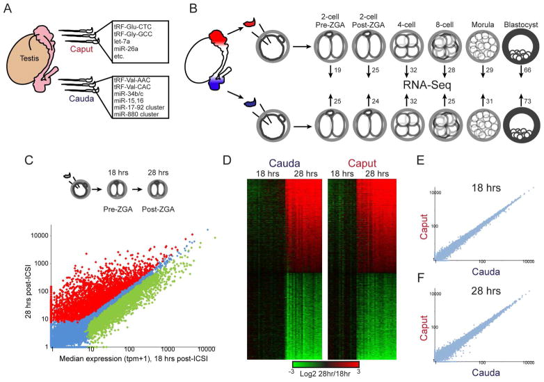
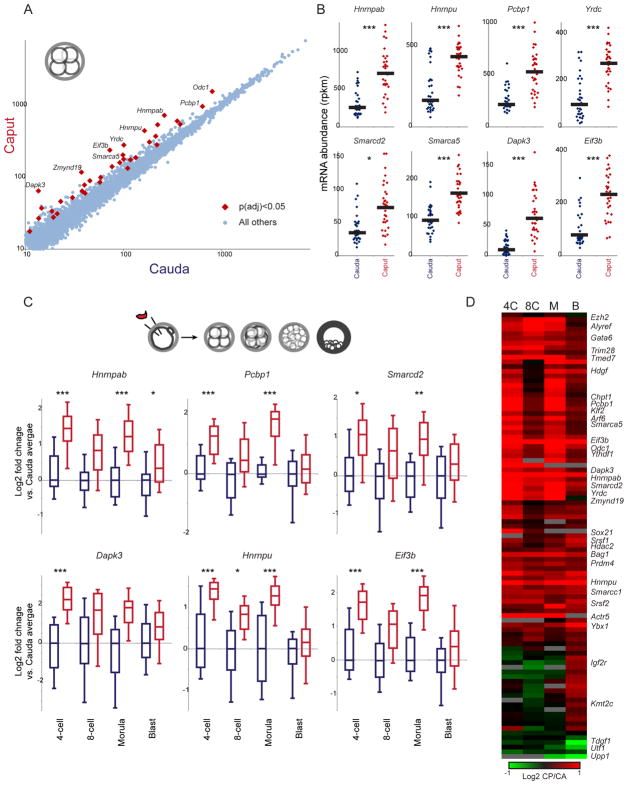
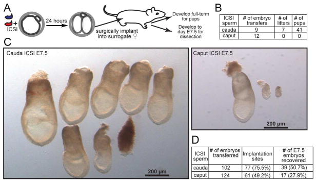
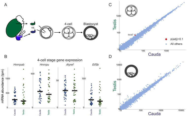
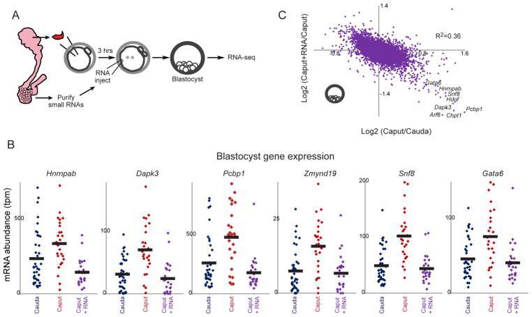
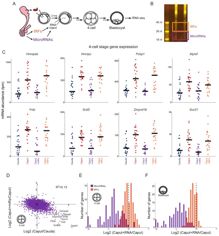
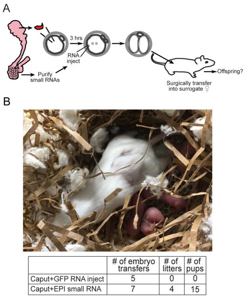
The small RNA payload of mammalian sperm undergoes dramatic remodeling during development, as several waves of microRNAs and tRNA fragments are shipped to sperm during post-testicular maturation in the epididymis. Here, we take advantage of this developmental process to probe the function of the sperm RNA payload in preimplantation development. We generated zygotes via intracytoplasmic sperm injection (ICSI) using sperm obtained from the proximal (caput) versus distal (cauda) epididymis and then characterized the development of the resulting embryos. Embryos generated using caput sperm significantly overexpress multiple regulatory factors throughout preimplantation development, subsequently implant inefficiently, and fail soon after implantation. Remarkably, microinjection of purified cauda-specific small RNAs into caput-derived embryos not only completely rescued preimplantation molecular defects but also suppressed the post-implantation embryonic lethality phenotype. These findings reveal an essential role for small RNA remodeling during post-testicular maturation of mammalian sperm and identify a specific preimplantation gene expression program responsive to sperm-delivered microRNAs.

Keywords: epigenetics; preimplantation development; small RNAs; spermatogenesis.


Conflict of interest statement

DECLARATION OF INTERESTS

The authors declare no competing interests.

Figures

Figure 1
Figure 1. Experimental pipeline
A) Sperm obtained from the proximal (caput) and distal (cauda) epididymis carry distinct small RNA populations (Nixon et al., 2015; Sharma et al., 2016). These sperm samples differ in the abundance of hundreds of small RNAs – shown here are prominent examples of relevant RNA species. The RNAs listed for caput sperm are present at high abundance in caput sperm and are generally maintained at high levels in cauda sperm, while those listed for cauda sperm are examples of small RNAs that are scarce in caput sperm but specifically gained in cauda sperm. See also Supplemental Figure S1. B) Intracytoplasmic sperm injection (ICSI) pipeline. Paired caput and cauda sperm samples (from the same animal) are injected into control oocytes, and embryos are cultured to the indicated stages and subject to single-embryo RNA-Seq. Numbers associated with each stage give the number of embryos in the initial baseline dataset (excluding later RNA injection datasets – see Table S7 for a complete listing of embryo numbers for the entire study). C) ICSI 2-cell stage dataset faithfully recapitulates the major wave of zygotic genome activation (ZGA). Scatterplot of average mRNA abundance (average tpm +1) for 18 hour embryos (pre-ZGA, x axis) vs. 28 hour embryos (post-ZGA, y axis), with significantly (adjusted p < 0.05) differentially-expressed genes shown as red and green dots. See also Supplemental Figure S2. D) Heatmaps of significant ZGA genes shown separately for caput- and cauda-derived embryos, as indicated. E–F) Scatterplots of average mRNA abundance (as in panel C) for Caput vs. Cauda embryos at 18 hours (E) and 28 hours (F), showing no significantly differentially-expressed genes.
Figure 2
Figure 2. Effects of post-testicular sperm maturation on preimplantation gene regulation
A) Scatterplot of 4-cell stage gene expression for Cauda vs. Caput embryos. Data are shown for all genes with a median expression level of at least 10 tpm across the entire dataset, and significant differences (here, showing 37 genes with t-test p value < 0.05, adjusted for multiple hypotheses) in gene expression are indicated. B) Data for every individual 4-cell embryo for eight genes overexpressed in Caput embryos. Black bars show median expression for each group of embryos. *, **, and *** show comparisons with adjusted p values below 0.1, 0.01, and 0.001, respectively. C) Gene expression differences at the 4-cell stage persist into later developmental stages. Here, box plots (box: median and 25th/75th percentiles, whiskers: 10th and 90th percentiles) show log2 of expression level normalized relative to the median value in matched cauda-derived embryos of the relevant developmental stage. D) Heatmap of all 95 genes exhibiting a difference in expression (padj < 0.1) at any stage of development. Data are expressed as log2 ratio (Caput/Cauda). See also Supplemental Figures S3–S4.
Figure 3
Figure 3. Caput-derived embryos fail during early post-implantation development
A) Schematic representation of embryo transfer experiments. ICSI embryos were cultured to the 2-cell stage, then 15–20 embryos were transferred into an oviduct of a pseudo-pregnant Swiss Webster female. B) Success rates for Caput and Cauda embryos allowed to develop to term. Note that Caput embryos were also transferred to recipient females in additional experiments below, with a total success rate of 0/20 across all experiments in this study. C) Images of all embryos dissected from females transferred with Cauda or Caput embryos. A greater fraction of Cauda embryos implanted and produced normally-developing embryos. The left panel shows six normal, and one abnormal, Cauda embryos. In contrast, fewer implantation sites were observed for the Caput embryos, and the few implantation sites observed were either undergoing resorption or carried abnormally developing embryos. The right panel shows one normal embryo and two abnormal ones. D) Quantitation of all embryo transfer dissections. Caput-derived embryos exhibit lower implantation rates, higher resorption rates, and (not quantified but shown in panel C) poor growth in the peri-implantation period. Note that percentages associated with number of E7.5 embryos recovered are relative to number of implantation sites, not total number of embryos transferred.
Figure 4
Figure 4. Molecular analysis of testicular sperm-derived preimplantation embryos
A) Schematic of testicular sperm ICSI experiments. B) Dot plots showing examples of key Caput-regulated genes in individual 4-cell stage Cauda (n=33) and Testicular (n=34) embryos. Black bars show median expression level for each condition. C–D) Scatterplots comparing mRNA abundance (all genes with median tpm > 10) between Cauda and Testicular embryos at the 4-cell (C) and blastocyst (D) stages. See also Supplemental Figure S5 for morula-stage data.
Figure 5
Figure 5. Cauda-specific small RNAs rescue development of caput-derived embryos
A) Schematic representation of cauda epididymosomal RNA purification and microinjection. ICSI zygotes were generated by injection of caput or cauda sperm. Three hours later, Caput embryos were microinjected either with gel-purified small (18–40 nt) RNAs obtained from cauda epididymosomes, along with H3.3-GFP mRNA as a tracer for successful injections, or were injected with H3.3-GFP alone. Resulting zygotes were cultured to the blastocyst stage for RNA-Seq. B) Cauda-specific RNAs repress genes overexpressed in caput-derived embryos. Dots show individual embryo RNA-Seq data for Cauda (n=39), Caput (n=26), or Caput+RNA (n=25) blastocysts. In every case, injection of cauda-specific small RNAs returns expression of these genes to the Cauda baseline. C) Scatterplot of Caput effects on mRNA abundance (x axis, log2 fold change Caput/Cauda) vs. effects of small RNA injection (y axis, log2 fold change Caput+RNA/Caput). Dots in lower right corner are overexpressed in Caput embryos, and downregulated upon microinjection of cauda-specific small RNAs.
Figure 6
Figure 6. Cauda-specific microRNAs, but not tRFs, rescue Caput preimplantation defects
A) Schematic representation of microinjection experiment. As in Figure 5A, but two separate small RNA populations were gel-purified – 18–26 nt RNAs are enriched for microRNAs (but include additional molecular species including short 5’ and 3’ tRFs), while 26–40 nt RNAs are primarily comprised of tRNA fragments. B) Purification of epididymosomal RNAs. Acrylamide electrophoresis of two replicate total RNA samples purified from cauda epididymosomes. Boxes show the boundaries used for gel-purification of microRNAs and tRFs. C) mRNA abundance for individual 4-cell stage Cauda (n=23), Caput (n=23), Caput+microRNA (n=22), and Caput+tRF (n=21) embryos, for representative Caput-upregulated genes. In all cases, cauda-specific “microRNAs” returned mRNA abundance to Cauda levels, while tRFs had little to no effect on these target genes. See Supplemental Figure S6B for blastocyst-stage data. The clear difference between these matched RNA injections – both RNA samples should be free of contaminating proteins, and should include similar levels of any potential contaminants such as leftover acrylamide – demonstrates that the rescue activity is specific to 18–26 nt cauda epididymosomal small RNAs. D) Scatterplot comparing Caput effects (x axis) and microRNA effects (y axis) on mRNA abundance in 4-cell stage embryos. See Supplemental Figure S6C for blastocyst stage dataset. E–F) Histograms of microRNA and tRF effects on Caput-upregulated genes. In both cases, data are shown for mRNAs which exhibit a log2 fold change of at least 0.5 in Caput vs. Cauda embryos (p<0.01, uncorrected for multiple hypothesis testing to include a greater number of genes – results are even more dramatic for the smaller subset of genes significant after multiple hypothesis-correction). For both 4-cell stage (E) and blastocyst-stage (F) data, cauda-specific microRNAs cause downregulation of Caput-induced genes, while tRFs have little to no effect on these genes (KS p value comparing microRNA and tRF effects < 10−15 for both datasets).
Figure 7
Figure 7. Cauda-specific small RNAs rescue development of Caput embryos
A) Experimental schematic. Small RNA injections were carried out as in Figure 5A, but ICSI embryos were transferred at the 2-cell stage into the oviduct of pseudo-pregnant females as in Figure 3. B) Image shows example of a successful litter generated from caput-derived embryos microinjected with cauda RNAs. Table underneath lists success rates, as in Figure 3B.

Comment in

References

    1. Arteaga-Vazquez MA, Chandler VL. Paramutation in maize: RNA mediated transgenerational gene silencing. Curr Opin Genet Dev. 2010;20:156–163. - PMC - PubMed
    1. Belleannee C, Calvo E, Caballero J, Sullivan R. Epididymosomes convey different repertoires of microRNAs throughout the bovine epididymis. Biology of reproduction. 2013;89:30. - PubMed
    1. Chen Q, Yan M, Cao Z, Li X, Zhang Y, Shi J, Feng GH, Peng H, Zhang X, Zhang Y, et al. Sperm tsRNAs contribute to intergenerational inheritance of an acquired metabolic disorder. Science (New York, N Y. 2016;351:397–400. - PubMed
    1. Donkin I, Versteyhe S, Ingerslev LR, Qian K, Mechta M, Nordkap L, Mortensen B, Appel EV, Jorgensen N, Kristiansen VB, et al. Obesity and Bariatric Surgery Drive Epigenetic Variation of Spermatozoa in Humans. Cell metabolism. 2016;23:369–378. - PubMed
    1. Feng S, Jacobsen SE, Reik W. Epigenetic reprogramming in plant and animal development. Science (New York, N Y. 2010;330:622–627. - PMC - PubMed

Publication types