Review
. 2018 Jun;8(3):253-271.
doi: 10.21037/cdt.2018.01.09.

Chronic pulmonary embolism: diagnosis

Affiliations
Review

Chronic pulmonary embolism: diagnosis

Katia Hidemi Nishiyama et al. Cardiovasc Diagn Ther. 2018 Jun.

Abstract

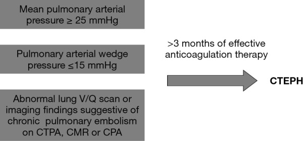
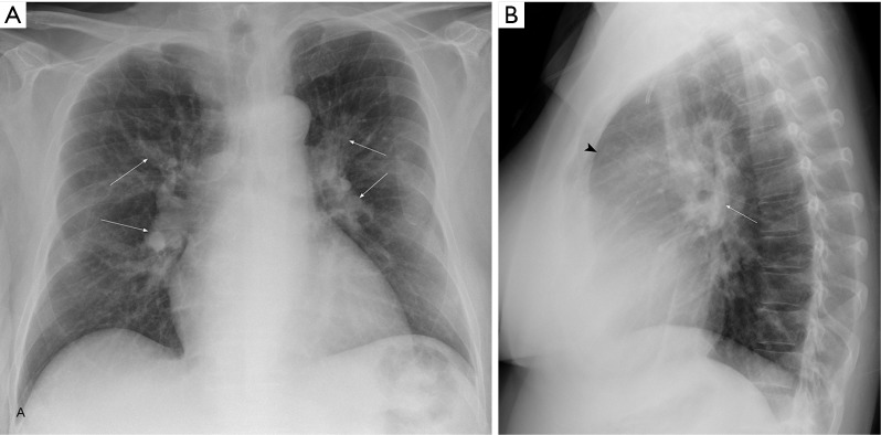
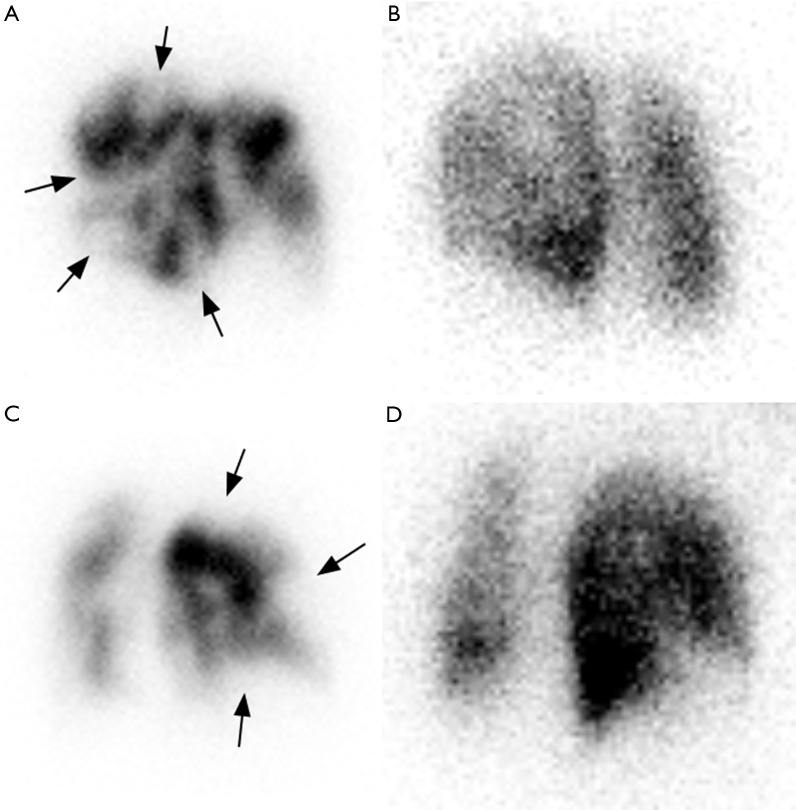
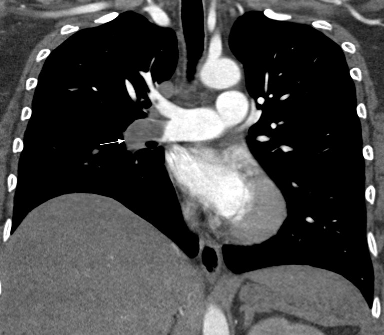
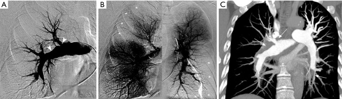
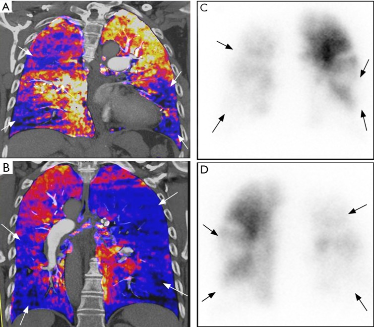
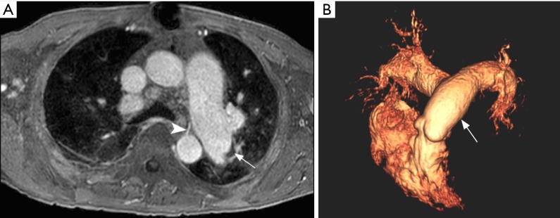
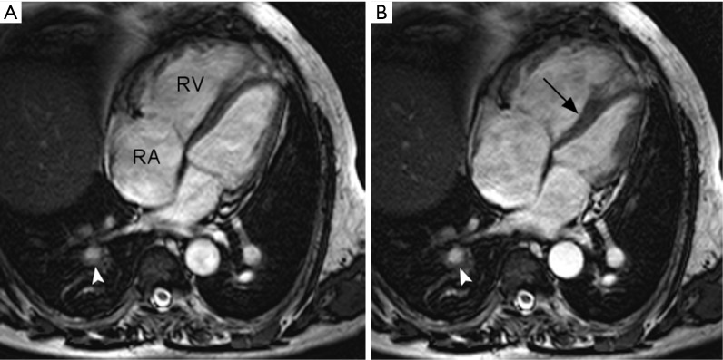
Chronic thromboembolic pulmonary hypertension (CTEPH) is a complication of venous thromboembolic disease. Differently from other causes of pulmonary hypertension, CTEPH is potentially curable with surgery (thromboendarterectomy) or balloon pulmonary angioplasty. Imaging plays a central role in CTEPH diagnosis. The combination of techniques such as lung scintigraphy, computed tomography and magnetic resonance angiography provides non-invasive anatomic and functional information. Conventional pulmonary angiography (CPA) with right heart catheterization (RHC) is considered the gold standard method for diagnosing CTEPH. In this review, we discuss the utility of these imaging techniques in the diagnosis of CTEPH.

Keywords: Pulmonary embolism; V/Q; chronic; computed tomography; pulmonary hypertension.


Conflict of interest statement

Conflicts of Interest: The authors have no conflicts of interest to declare.

Figures

Figure 1
Figure 1
Diagnostic criteria for chronic thromboembolic pulmonary hypertension. CTEPH, chronic thromboembolic pulmonary hypertension; lung V/Q scan, ventilation/perfusion lung scintigraphy; CTPA, computed tomography pulmonary angiography; CMR, cardiovascular magnetic resonance; CPA, conventional pulmonary angiography.
Figure 2
Figure 2
Frontal (A) and lateral (B) view chest radiographs show enlarged central pulmonary arteries (arrows), increased cardiothoracic index and obliteration of the retrosternal clear space (arrowhead).
Figure 3
Figure 3
Left posterior oblique (LPO) imaging with intravenous injection of albumin macroaggregate-99mTc revealing multiple segmental perfusion defects in the left lung (arrows, A), with corresponding normal ventilation on LPO imaging obtained with inhalation of 133Xenon (B). Right posterior oblique (RPO) perfusion scintigraphy also revealing mismatched segmental perfusion defects in the right lung (arrows, C) with normal ventilation (D).
Figure 4
Figure 4
Coronal CT pulmonary angiography image shows a larger thrombus in the right interlobar artery with complete occlusion and vessel cutoff (arrow).
Figure 5
Figure 5
CT pulmonary angiography appearances of chronic thrombi in different patients. Axial images (A) and (B) show webs (arrows), reflecting non-resolved thrombi and partial recanalization. Coronal image (C) shows an eccentric thrombus in right interlobar pulmonary artery with calcified mural thickening (arrow).
Figure 6
Figure 6
CT pulmonary angiography features of chronic thromboembolic pulmonary hypertension. Axial image (A) showing reduced caliber vessels (arrows) related to the contraction of the organizing thrombus and poor contrast opacification in comparison with contralateral arteries (arrowheads). Axial maximum intensity projection (MIP) at the thoracoabdominal transition (B) showing a prominent phrenic artery (arrow) providing systemic collateral circulation, in addition to asymmetric caliber of the pulmonary arteries in the lower lobes (attenuated on the right side, arrowheads). Sagittal MIP reconstruction (C) reveals a prominent bronchial artery providing systemic collateral circulation (arrow).
Figure 7
Figure 7
CT evaluation of pulmonary hypertension. (A) Noncontrast axial CT shows main pulmonary artery (MPA) of 40 mm and MPA/aorta ratio > 1.1:1, consistent with pulmonary hypertension. The absence of contrast facilitates the observation of chronic eccentric thrombi with mural calcification (arrows). Axial CT pulmonary angiography image (B) shows right ventricular enlargement (right-to-left ventricular ratio greater than 1:1). Webs noted in right lower lobe arteries, consistent with chronic pulmonary embolism (arrow).
Figure 8
Figure 8
Regions of increased (arrowheads) and decreased lung attenuation (arrows) characterize lung mosaicism. Markedly reduced diameter of segmental/subsegmental arteries noted within areas of decreased lung attenuation.
Figure 9
Figure 9
Conventional pulmonary angiography correlated with CT pulmonary angiography (CTPA). Selective right pulmonary angiography (A) showing irregularities at the origin of the right upper lobar and proximal interlobar pulmonary arteries (arrows in A), suggestive of chronic pulmonary embolism. Composite image of right and left pulmonary angiography (B) during parenchymal enhancement shows multiple hypoperfused areas, especially in right upper and left lower lobes (asterisks). Coronal maximum intensity projection CTPA (C) shows arterial wall thickening and luminal narrowing in the right pulmonary artery and segmental branches of the left lower lobar pulmonary artery (arrows).
Figure 10
Figure 10
Dual-energy CT with V/Q scan correlation. (A,B) Coronal perfused blood volume map shows multiple segmental and lobar perfusion defects (blue areas, arrows). Planar anterior (C) and posterior (D) perfusion scintigraphy shows corresponding perfusion defects with similar distribution.
Figure 11
Figure 11
Magnetic resonance pulmonary angiography (MRPA). Axial post contrast fat-suppressed spoiled gradient recalled echo (A) shows left pulmonary artery dilation with a subtle eccentric crescent-shaped thrombus (arrow). Note a prominent bronchial artery arising from the descending aorta (arrowhead). Tridimensional volume rendered reconstruction (B) showing dilation of the main pulmonary artery (arrowhead) and central branches.
Figure 12
Figure 12
Cine balanced steady-state free precession imaging. Four-chamber view in end-diastole (A) and end-systole (B) showing dilation of the right atrium (RA) and ventricle (RV). Note paradoxical deviation of the interventricular septum towards the left (arrow) during systole. Pulmonary artery wall thickening and luminal narrowing is also seen in the right lower lobe (arrowheads), consistent with sequelae of chronic thromboembolic pulmonary hypertension (CTEPH).
Figure 13
Figure 13
CT pulmonary angiography (CTPA) images in acute pulmonary embolism. Axial CTPA images (A and B) show partial filling defects, centrally located in the right pulmonary artery (arrow) and eccentric in segmental branches (arrowheads). Note acute angle between thrombi and vessel walls. Axial lung window (C) shows “reversed halo sign” in the right lower lobe (arrows), a nonspecific sign of pulmonary infarction.
Figure 14
Figure 14
CT pulmonary angiography images in partially occlusive chronic pulmonary embolism. Note peripheral crescent-shaped defects presenting an obtuse angle with vessel wall (arrows in A and B) in contrast with acute angle observed in acute embolism (Figure 13).

References

    1. Beckman MG, Hooper WC, Critchley SE, et al. Venous thromboembolism: a public health concern. Am J Prev Med 2010;38:S495-501. 10.1016/j.amepre.2009.12.017 - DOI - PubMed
    1. Pengo V, Lensing AW, Prins MH, et al. Incidence of chronic thromboembolic pulmonary hypertension after pulmonary embolism. N Engl J Med 2004;350:2257-64. 10.1056/NEJMoa032274 - DOI - PubMed
    1. Ende-Verhaar YM, Cannegieter SC, Vonk Noordegraaf A, et al. Incidence of chronic thromboembolic pulmonary hypertension after acute pulmonary embolism: a contemporary view of the published literature. Eur Respir J 2017;49. pii: 1601792. 10.1183/13993003.01792-2016 - DOI - PubMed
    1. Lang IM, Pesavento R, Bonderman D, et al. Risk factors and basic mechanisms of chronic thromboembolic pulmonary hypertension: a current understanding. Eur Respir J 2013;41:462-8. 10.1183/09031936.00049312 - DOI - PubMed
    1. Humbert M. Pulmonary arterial hypertension and chronic thromboembolic pulmonary hypertension: pathophysiology. Eur Respir Rev 2010;19:59-63. 10.1183/09059180.00007309 - DOI - PMC - PubMed

LinkOut - more resources