Review
. 2018 Jun;8(3):338-349.
doi: 10.21037/cdt.2017.12.08.

Pulmonary arteriovenous malformations: endovascular therapy

Affiliations
Review

Pulmonary arteriovenous malformations: endovascular therapy

Murthy R Chamarthy et al. Cardiovasc Diagn Ther. 2018 Jun.

Abstract

Pulmonary arteriovenous malformations (PAVM) are abnormal direct communications between the branches of pulmonary arteries and veins, and are often seen in patients with hereditary hemorrhagic telangiectasia (HHT). If untreated, the right to left shunt can result in symptoms of hypoxemia, paradoxical emboli to the left side circulation, stroke and intracranial abscess. Endovascular therapy is a minimally invasive outpatient based treatment wherein the feeding artery to the PAVM is occluded with coils or plugs or a combination of both and is associated with minimal morbidity and no mortality. In this manuscript, we will review the indications and contraindications for endovascular therapy, pre-procedural work up, procedure technique and variations, complications, and outcomes.

Keywords: Pulmonary arteriovenous malformation (PAVM); coil; embolization; hereditary hemorrhagic telangiectasia (HHT); occlusion; plug.


Conflict of interest statement

Conflicts of Interest: The authors have no conflicts of interest to declare.

Figures
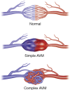
Figure 1
Figure 1
Pulmonary AVM illustrations. A normal capillary network is noted between the pulmonary arterial branch and pulmonary venous tributary facilitating functions of gaseous exchange and filtration. A single segmental arterial feeder connects directly to the draining vein through the nidus or sac without intervening capillary network in a simple PAVM. Multiple segmental feeding arterial branches are identified in a complex PAVM. PAVM, pulmonary arteriovenous malformations.
Figure 2
Figure 2
Types of pulmonary AVM. (A) Demonstrates a simple left lower lobe PAVM with a single arterial feeder (red arrow) and a draining vein (blue arrow). Multiple segmental feeders are noted in a complex left lower lobe PAVM (B, red arrow). CT scan (C) and left pulmonary arteriogram (D) demonstrate findings of diffuse PAVM in a patient with HHT. PAVM, pulmonary arteriovenous malformations; HHT, hereditary hemorrhagic telangiectasia.
Figure 3
Figure 3
Catheters used in pulmonary angiography and pulmonary arterial embolization. A wire and pigtail catheter can be utilized to gain access to the pulmonary artery. The right-angle shape of the angled pigtail catheter (preferred catheter) allows easy access from the right atrium to the right ventricle and further selects the pulmonary arteries. Lumax system (7 Fr guiding catheter with a 5 Fr catheter combination) allows easy selection of pulmonary arterial branches and provides stable access for embolization.
Figure 4
Figure 4
An illustration of various embolic occlusion devices and techniques. Coil, Amplatzer type IV plug and microvascular plug occlusion techniques are illustrated in images (A,B,C) respectively; (D) illustrates combination techniques with additional coil embolization following Amplatzer or microvascular plug deployment. Arrows demonstrate blood flow direction within the pulmonary arterial branch feeding the pulmonary arteriovenous malformation.
Figure 5
Figure 5
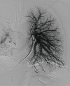
Coil embolization. In this patient with HHT and multiple PAVM, left pulmonary arteriogram demonstrates complete occlusion of the PAVM with coil embolization alone. PAVM, pulmonary arteriovenous malformations.
Figure 6
Figure 6
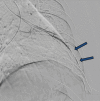
Embolization with Amplatzer vascular plug. A left lower lobe complex PAVM (Figure 2B) was successful occluded with Amplatzer type 4 vascular plugs (blue arrows). PAVM, pulmonary arteriovenous malformations.
Figure 7
Figure 7
Embolization with Amplatzer vascular plug and coils. Persistent perfusion of the left lower lobe PAVM was noted after embolization with Amplatzer type 4 vascular plug (A, red arrows). Additional coil embolization was performed proximal to the vascular plugs resulting in complete occlusion (B, blue arrow). PAVM, pulmonary arteriovenous malformations.
Figure 8
Figure 8
Occlusion with MVP plug. The feeder vessel of a left lower lobe simple PAVM (Figure 2A) was embolized with two MVP-3 vascular plugs resulting in complete occlusion (blue arrows). PAVM, pulmonary arteriovenous malformations.
Figure 9
Figure 9
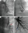
Embolization with coils and MVP plug. (A) Demonstrates persistent perfusion of a right lower lobe PAVM after coil embolization (red arrow). Additional MVP plug embolization results in complete occlusion (B, blue arrow). PAVM, pulmonary arteriovenous malformations.
Figure 10
Figure 10
Occlusion with MVP plug and coils. Embolization with MVP plug and coils (yellow and blue arrows respectively in A) resulted in successful occlusion of the right lower lobe PAVM seen on pretreatment (B). PAVM, pulmonary arteriovenous malformations.
Figure 11
Figure 11
Adjunctive role of stent-graft placement. Left pulmonary arteriogram and a selective left lower lobe branch arteriogram demonstrate a PAVM (A and B respectively). Arteriogram post embolization with a MVP-7 vascular plug (C) demonstrates an additional tiny tortuous feeder not feasible for catheterization and embolization (red arrow). A stent-graft (5 mm × 16 mm iCast, blue arrow) was deployed within the lower lobe pulmonary arterial branch successfully excluding the PAVM feeder (D). PAVM, pulmonary arteriovenous malformations.
Figure 12
Figure 12
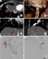
Embolization of a recanalized AVM with MVP vascular plug. CT (A) and angiography (B) demonstrate recanalization (red arrows) of a previously embolized left lower lobe PAVM. (C) Demonstrates successful occlusion with a MVP plug (blue arrow). PAVM, pulmonary arteriovenous malformations.
Figure 13
Figure 13
Occlusion of a recanalized AVM with dense coil packing. CT (A) and selective pulmonary branch arteriogram (B) demonstrate recanalization (red arrows) of a previously embolized right lower lobe PAVM. (C) Demonstrates successful occlusion with dense coil packing (blue arrow). PAVM, pulmonary arteriovenous malformations.
Figure 14
Figure 14
Reperfusion from systemic artery. CT images (axial A, and sagittal B and C) demonstrate reperfusion of a previously embolized right lower lobe PAVM via systemic feeder branches from internal mammary collaterals (red arrows). PAVM, pulmonary arteriovenous malformations.
Figure 15
Figure 15
Reperfusion from systemic artery. 3D CT images (A, red arrows) demonstrate systemic feeders to the previously embolized right lower lobe PAVM from right phrenic artery. Selective phrenic arteriogram confirms the findings seen on CT (B, red arrow). The feeder branch from phrenic artery was successfully embolized with coils (C, blue arrow). PAVM, pulmonary arteriovenous malformations.

References

    1. Shovlin CL, Jackson JE, Bamford KB, et al. Primary determinants of ischaemic stroke/brain abscess risks are independent of severity of pulmonary arteriovenous malformations in hereditary haemorrhagic telangiectasia. Thorax 2008;63:259-66. 10.1136/thx.2007.087452 - DOI - PubMed
    1. Pollak JS, Saluja S, Thabet A, et al. Clinical and anatomic outcomes after embolotherapy of pulmonary arteriovenous malformations. J Vasc Interv Radiol 2006;17:35-44; quiz 5. 10.1097/01.RVI.0000191410.13974.B6 - DOI - PubMed
    1. Pierucci P, Murphy J, Henderson KJ, et al. New definition and natural history of patients with diffuse pulmonary arteriovenous malformations: twenty-seven-year experience. Chest 2008;133:653-61. 10.1378/chest.07-1949 - DOI - PubMed
    1. Zukotynski K, Chan RP, Chow CM, et al. Contrast echocardiography grading predicts pulmonary arteriovenous malformations on CT. Chest 2007;132:18-23. 10.1378/chest.06-2356 - DOI - PubMed
    1. Ference BA, Shannon TM, White RI, Jr, et al. Life-threatening pulmonary hemorrhage with pulmonary arteriovenous malformations and hereditary hemorrhagic telangiectasia. Chest 1994;106:1387-90. 10.1378/chest.106.5.1387 - DOI - PubMed

LinkOut - more resources