Review
. 2018 Jun;8(3):362-371.
doi: 10.21037/cdt.2018.03.03.

Post-operative imaging of pulmonary vessels

Affiliations
Review

Post-operative imaging of pulmonary vessels

Carlos Santiago Restrepo et al. Cardiovasc Diagn Ther. 2018 Jun.

Abstract

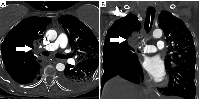
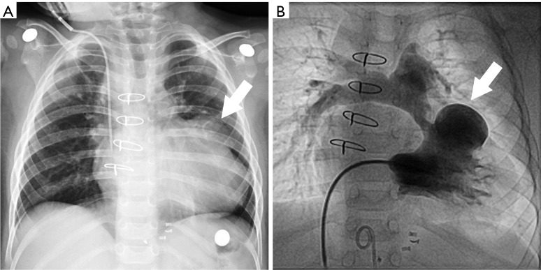
Complications following cardiothoracic surgery are responsible for prolonged hospital stay, increase cost in patient care and increased morbidity and mortality. Vascular complications in particular are significant contributors to poor patient outcome due to either hemorrhage or thrombosis and ischemia. Evaluation of vascular complications in the postoperative patient requires a rapid and reliable imaging approach. Vascular complications after cardiothoracic surgery include pulmonary artery thrombosis, pseudoaneurysm, pulmonary vein thrombosis, vascular fistulas, stenosis and infarction. Multidetector CT (MDCT), often the imaging modality of choice, offers a one-stop-shop capability to visualize the entire cardiothoracic vasculature, airways, lung parenchyma, mediastinum and chest wall with excellent temporal and spatial resolution.

Keywords: Pulmonary circulation; X-ray computed; pulmonary artery; pulmonary veins; thoracic surgery; tomography.


Conflict of interest statement

Conflicts of Interest: The authors have no conflicts of interest to declare.

Figures

Figure 1
Figure 1
Pulmonary artery pseudoaneurysm in a 70-year-old patient status post right upper lobectomy for lung cancer, who presents with hemoptysis. (A) Contrast enhanced CT demonstrates a pseudoaneurysm in the posterior aspect of the right hilum (arrow); (B) digital subtraction angiography, selective injection in the right pulmonary artery confirms the pseudoaneurysm at the resection site (arrow).
Figure 2
Figure 2
Pulmonary artery mismatch in a 63-year-old female patient status post bilateral lung transplant. Contrast enhanced CT shows the discrepancy in size between the recipient and donor pulmonary arteries with a step-off configuration at the level of the anastomosis (arrow).
Figure 3
Figure 3
Right pulmonary artery stenosis in a 61-year-old male patient status post lung transplant. Digital subtraction angiography confirms a “waist” with luminal narrowing at the right pulmonary artery anastomosis (arrow).
Figure 4
Figure 4
Post lung transplant pulmonary artery thrombosis with pulmonary infarction. Contrast enhanced CT axial image (A) and coronal reconstruction (B) demonstrate complete obstruction of the left pulmonary artery with collapse of the entire left lung.
Figure 5
Figure 5
Pulmonary vein thrombosis in a 57-year-old female patient with single left lung transplantation. Contrast enhanced CT axial image shows filling defect in the anastomotic site between the left atrium and left lower lobe pulmonary vein (arrow).
Figure 6
Figure 6
Systemic artery to pulmonary artery fistula in a 52-year-old female patient with history of revascularization surgery (CABG) with several complications that required surgical reintervention. (A) Contrast enhanced CT shows a tortuous intrapulmonary vessel in the anterior left upper lobe between the left internal mammary artery and the left axillary artery (arrow); (B) axial image further down reveal communication to the left pulmonary artery (arrow); (C,D) digital subtraction catheter angiography with selective injection in the left subclavian artery confirms the presence of fistulous arterial communications between the left side systemic arterial and pulmonary arterial circulation. CABG, coronary artery bypass graft.
Figure 7
Figure 7
Coronary-to-pulmonary artery fistula after coronary artery bypass graft. (A) Cardiac gated coronary artery CT axial image shows a high density small irregular vessel on the left lateral aspect of the pulmonary trunk (arrow); (B) coronal image shows the proximity between the tortuous irregular vessel and the left anterior descending (LAD) coronary artery (arrow); (C) sagittal maximum intensity projection (MIP); (D) volume rendered 3D reconstruction. Images show the connection between the fistula on the surface of the pulmonary trunk and the left anterior descending coronary artery (arrows). Arterial grafts to the circumflex and LAD are also appreciated.
Figure 8
Figure 8
Lobar torsion in a 66-year-old female patient status post right upper lobectomy for lung cancer. A few days later her status worsens. (A) Contrast enhanced CT axial image; (B) contrast enhanced CT coronal reconstruction. Images demonstrate mass-like opacity of the right hilum with interruption of the right pulmonary artery, interruption of the right superior pulmonary vein and narrowing of the right middle lobe bronchi (arrows). Patient was taken to surgery and found to have devascularization of the right lung with torsion of the right middle lobe.
Figure 9
Figure 9
Post-lobectomy pulmonary artery thrombosis. Contrast enhanced CT, coronal reconstruction in a patient with right lower lobe and middle lobe resection demonstrates right pulmonary artery stump thrombosis (arrows).
Figure 10
Figure 10
Post-pneumonectomy pulmonary embolism. Contrast enhanced axial image at the level of the left hilum shows extensive filling defect in the left pulmonary artery (arrow) in this patient with previous right side pneumonectomy.
Figure 11
Figure 11
TOF post RVOT repair with pseudoaneurysm at the suture line. (A) Chest radiograph shows an abnormal round mass-like opacity projecting on top of the left hilum (arrow); (B) contrast enhanced CT reveals a pseudoaneurysm of the anterolateral aspect of the right ventricular outflow tract (arrow). TOF, tetralogy of Fallot; RVOT, right ventricular outflow tract.
Figure 12
Figure 12
RVOT pseudoaneurysm in a 15-month-old girl status post TOF repair. (A) Chest radiograph shows an abnormal bulge on the lateral aspect of the cardiac silhouette (arrow); (B) digital subtraction angiography with right ventricular injection confirms pseudoaneurysm of the RVOT (arrow) with severe pulmonary artery stenosis. TOF, tetralogy of Fallot; RVOT, right ventricular outflow tract.
Figure 13
Figure 13
Pulmonary artery stenosis in two different patients, status post repaired TOF. (A) A 22-year-old male, contrast enhanced CT shows long segment stenosis of the left pulmonary artery with surgical changes in the pulmonary trunk and proximal left pulmonary artery (arrow); (B) a 22-year-old male, contrast enhanced CT shows a pseudoaneurysm of the pulmonary trunk (large arrow) with distal stenosis (small arrow). TOF, tetralogy of Fallot.

References

    1. Petterfy A, Henze A. Hemorrhagic complications during pulmonary resection: a retrospective review of 1428 resections with 113 haemorrhagic episodes. Scand J Thorac Cardiov Surg 1983;17:283-7. 10.3109/14017438309099366 - DOI - PubMed
    1. Sirbu H, Busch T, Aleksic I, et al. Chest re-exploration for complications after lung surgery. Thorac Cardiovasc Surg 1999;47:73-6. 10.1055/s-2007-1013114 - DOI - PubMed
    1. Krasna MJ, Deshmukh S, McLaughlin JS. Complications of thoracoscopy. Ann Thorac Surg 1996;61:1066-9. 10.1016/0003-4975(96)00021-5 - DOI - PubMed
    1. Ziamik E, Grogan EL. Post-lobectomy early complications. Thorac Surg Clinics 2015;25:355-64. 10.1016/j.thorsurg.2015.04.003 - DOI - PMC - PubMed
    1. Restrepo CS, Carswell AP. Aneurysms and pseudoaneurysms of the pulmonary vasculature. Semin Ultrasound CT MR 2012;33:552-66. 10.1053/j.sult.2012.04.001 - DOI - PubMed

LinkOut - more resources