Review
. 2018 Jun;8(3):387-398.
doi: 10.21037/cdt.2018.05.05.

Radiologic review of acquired pulmonary vein stenosis in adults

Affiliations
Review

Radiologic review of acquired pulmonary vein stenosis in adults

Mauricio Galizia et al. Cardiovasc Diagn Ther. 2018 Jun.

Abstract

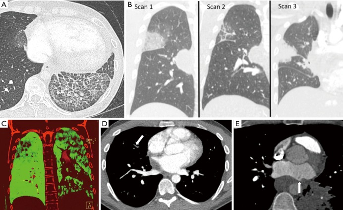
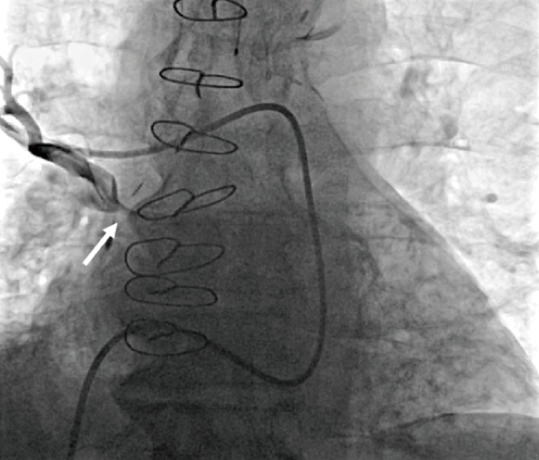
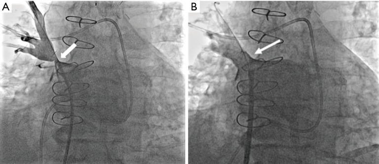
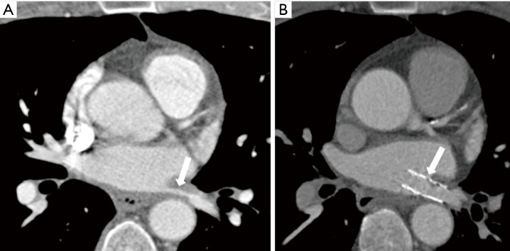
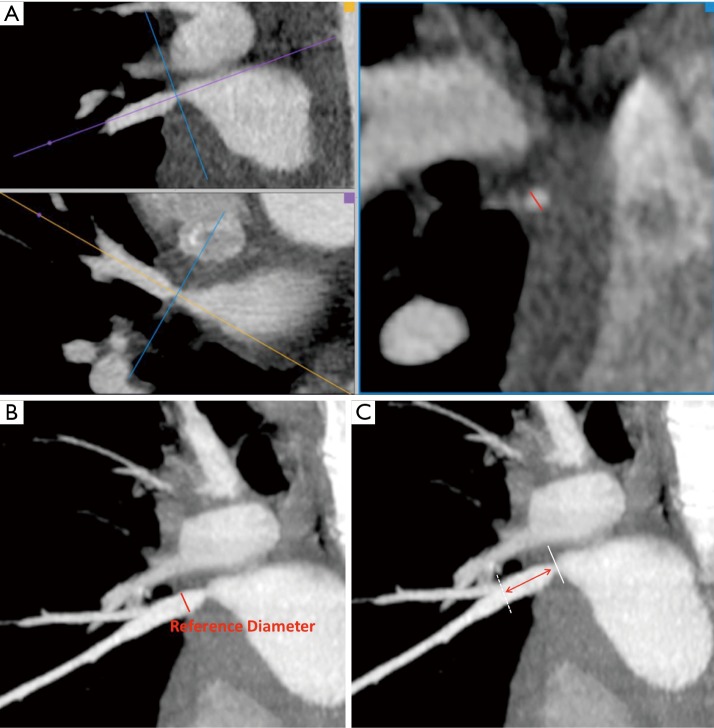
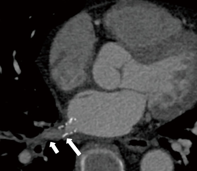
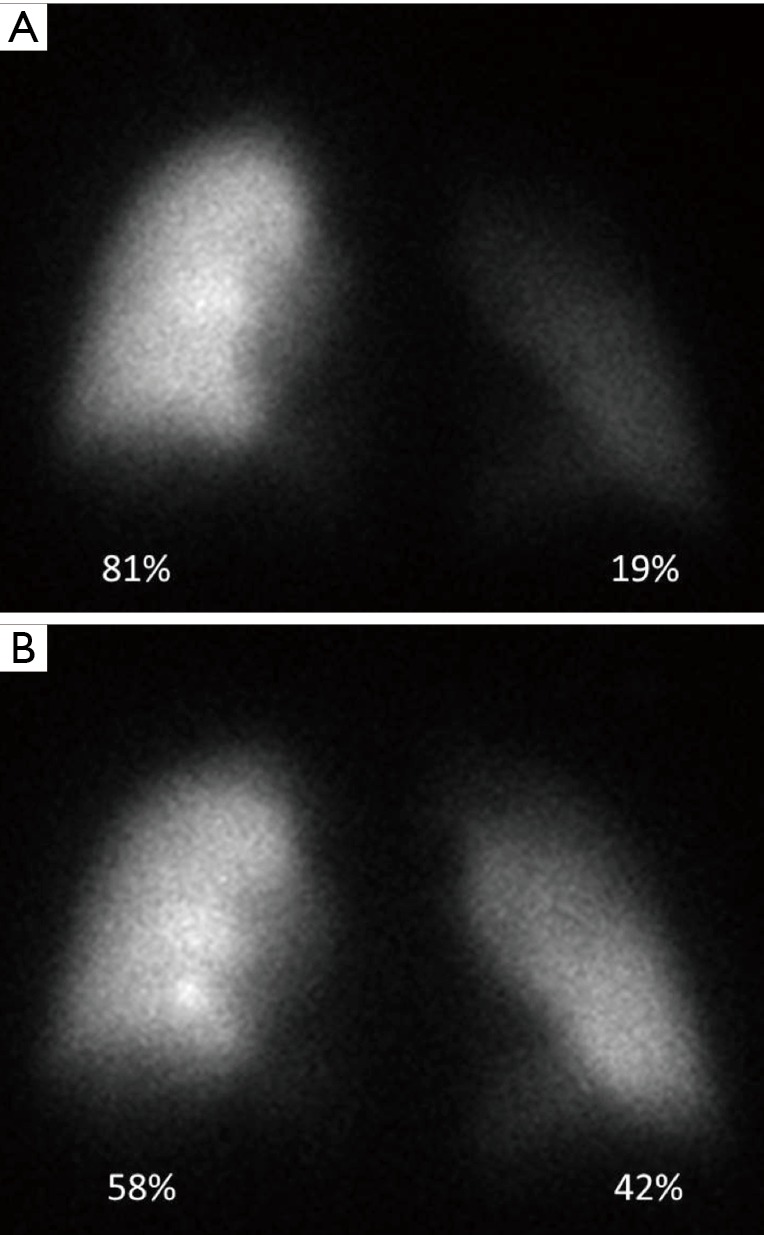
Acquired pulmonary vein stenosis (PVS) is an uncommon occurrence in adults, but one that carries significant morbidity/mortality. PVS can be secondary to neoplastic infiltration/extrinsic compression, non-neoplastic infiltration/extrinsic compression, or iatrogenic intervention. This article: (I) reviews the common causes of acquired PVS; (II) illustrates direct and indirect cross-sectional imaging findings in acquired PVS (in order to avoid misinterpretation of these imaging findings); and (III) details the role of imaging before and after the treatment of acquired PVS.

Keywords: Pulmonary vein stenosis (PVS); computed tomography (CT); imaging; magnetic resonance imaging (MRI); pulmonary vein ablation; pulmonary vein isolation (PVI); radiology.


Conflict of interest statement

Conflicts of Interest: The authors have no conflicts of interest to declare.

Figures

Figure 1
Figure 1
Embryologic development of the pulmonary veins. (A) Illustration shows that in early gestation, the veins draining the primitive lung buds do not connect to the primitive left atrium, but rather connect to various systemic venous systems (e.g., cardinal venous system, umbilical venous system) through the pulmonary venous plexus; the primitive pulmonary vein appears as an outpouching of the left atrium; (B) illustration shows that after 30 days of gestation the primitive pulmonary vein enlarges and connects to the pulmonary venous plexus, establishing for the first time venous flow from the lung buds to the left atrium; the connections of the pulmonary plexus with the systemic veins atrophy.
Figure 2
Figure 2
Diagram demonstrating the normal anatomic variants of the pulmonary veins. L1, two separate ostia for the left pulmonary veins that are not separated by atrial wall; L2, two separate ostia for the left pulmonary veins that are separated by atrial wall; L3, the left lower PV drains into the left upper PV with a short (<1 cm) common segment; L4, the left lower PV drains into the left upper PV with a long (>1 cm) common segment; R1, two separate ostia for the right pulmonary veins, with the vein draining the RML joining the right upper PV and forming a short (<1 cm) common segment; R2, three separate ostia for the right-sided pulmonary veins; R3, two separate ostia for the right pulmonary veins, with the vein draining the RML joining the right upper PV and forming a long (>1 cm) common segment; R4, two separate ostia for the right pulmonary veins, with the vein draining the RML joining the right lower PV; R5, four separate ostia for the right-sided pulmonary veins, with two veins draining the RML; R6, common ostium for the right-sided pulmonary veins; R7, four separate ostia for the right-sided pulmonary veins, with a separate ostium for the vein draining the RML and an accessory right top PV that drains the apical segment of the RUL. The most common anatomic pattern is L1/R1, with a total of four pulmonary veins draining into the left atrium. LUL, left upper lobe; LLL, left lower lobe; RUL, right upper lobe; RML, right middle lobe; RLL, right lower lobe; RTPV, right top pulmonary vein; PV, pulmonary vein.
Figure 3
Figure 3
Acquired pulmonary vein stenosis in adults secondary to lymphoma, fibrosing mediastinitis, bronchogenic cyst, and percutaneous radiofrequency catheter ablation for atrial fibrillation. (A) Axial reconstructed maximal intensity projection CT image shows lymphoma demonstrating mass effect on the left superior pulmonary vein with resultant stenosis (arrow); (B) axial reconstructed CT image shows fibrosing mediastinitis demonstrating mass effect on the right superior pulmonary vein with resultant stenosis (arrow); (C) coronal CT image shows bronchogenic cyst demonstrating mass effect on the right superior pulmonary vein with resultant stenosis (arrow); (D) coronal reconstructed maximal intensity projection MR image shows right inferior pulmonary vein stenosis after percutaneous radiofrequency catheter ablation for atrial fibrillation (arrow).
Figure 4
Figure 4
Direct cross-sectional imaging findings of acquired pulmonary vein stenosis in an adult. (A) Axial reconstructed maximal intensity projection CT image shows abrupt cut-off of the left superior pulmonary vein (arrow); (B) axial reconstructed CT image shows focal narrowing of the right inferior pulmonary vein (arrow).
Figure 5
Figure 5
Indirect cross-sectional imaging findings of acquired pulmonary vein stenosis in an adult: lung edema, waxing and waning lung opacities, decreased perfusion to the lung that is drained by the stenosed vein, non-opacified peripheral pulmonary veins, and mixing artifact at the interface between the stenosed pulmonary vein and the left atrium. (A) Axial CT shows interlobular septal thickening, intralobular interstitial thickening, and ground-glass opacity in the left lower lobe; lung edema secondary to left inferior pulmonary vein stenosis (can be mistaken for lung infection or lung hemorrhage); (B) coronal CT images from serial CT scans demonstrate waxing and waning lung opacities in the right upper and right middle lobes secondary to right superior pulmonary vein stenosis (can be mistaken for lung infection or lung infarction from pulmonary embolism); (C) coronal dual-energy CT image in demonstrates decreased lung perfusion to the left upper, left lower, and right upper lobes secondary to left superior pulmonary vein stenosis, left inferior pulmonary vein stenosis, right superior pulmonary vein stenosis; (D) axial CT image shows nonopacification of the right upper lobe pulmonary veins secondary to right superior pulmonary vein stenosis (can be mistaken for pulmonary emboli); (E) axial CT image shows mixing artifact at the interface between the left superior pulmonary vein and the left atrium (arrow) secondary to left superior pulmonary vein stenosis (can be mistaken for thrombus).
Figure 6
Figure 6
Pulmonary artery wedge angiography. Fluoroscopic angiographic image obtained with the catheter wedged in a right upper lobe pulmonary artery branch shows right superior pulmonary vein stenosis (arrow).
Figure 7
Figure 7
Direct pulmonary venography. (A) Fluoroscopic angiographic image obtained during a direct pulmonary venography before stenting shows right superior pulmonary vein stenosis (arrow); (B) fluoroscopic angiographic image obtained during a direct pulmonary venography after stenting shows elimination of the right superior pulmonary vein stenosis (arrow).
Figure 8
Figure 8
Percutaneous intervention for acquired pulmonary vein stenosis in an adult secondary to percutaneous radiofrequency catheter ablation for atrial fibrillation. (A) Axial reconstructed CT image shows left superior pulmonary vein stenosis before percutaneous intervention (arrow); (B) axial reconstructed CT image shows increased pulmonary vein diameter after percutaneous stent placement (arrow).
Figure 9
Figure 9
Planning a stenting procedure for pulmonary vein stenosis treatment by using contrast-enhanced CT multiplanar reconstruction. (A) Measurement of the minimum diameter at the stenosis in a true perpendicular plane (red line); (B) measurement of the reference diameter, the maximum diameter of the pulmonary vein distal to the stenosis (red line); (C) measurement of the distance from the stenosis site to the first distal branching point (red double arrow).
Figure 10
Figure 10
In-stent restenosis after percutaneous stenting of a right inferior pulmonary vein stenosis secondary to fibrosing mediastinitis. Axial reconstructed CT image demonstrates hypoattenuating material in the distal half of the stent (long arrow), suggestive of intimal proliferation, and poor contrast opacification of the pulmonary vein segment distal to the stent (short arrow), suggesting in-stent restenosis.
Figure 11
Figure 11
Role of quantitative lung perfusion scintigraphy before and after percutaneous stenting of a left inferior pulmonary vein stenosis in an adult secondary to percutaneous radiofrequency catheter ablation for atrial fibrillation. (A) Pre-treatment quantitative lung perfusion scintigraphy demonstrates decreased perfusion of the left lung as compared to the right lung; (B) post stenting quantitative lung perfusion scintigraphy demonstrates improved perfusion to the left lung when compared to the pre-treatment study.

References

    1. Sun CC, Doyle T, Ringel RE. Pulmonary vein stenosis. Hum Pathol 1995;26:880-6. 10.1016/0046-8177(95)90011-X - DOI - PubMed
    1. Latson LA, Prieto LR. Congenital and acquired pulmonary vein stenosis. Circulation 2007;115:103-8. 10.1161/CIRCULATIONAHA.106.646166 - DOI - PubMed
    1. Neill CA. Development of the pulmonary veins with reference to the embryology of anomalies of pulmonary venous return. Pediatrics 1956;18:880-7. - PubMed
    1. Marom EM, Herndon JE, Kim YH, et al. Variations in pulmonary venous drainage to the left atrium: implications for radiofrequency ablation. Radiology 2004;230:824-9. 10.1148/radiol.2303030315 - DOI - PubMed
    1. Can MM. Severe pulmonary vein stenosis due to invasion of metastatic lung cancer. Anadolu Kardiyol Derg 2013;13:E17. - PubMed