. 2018 Aug 31;128(9):3779-3793.
doi: 10.1172/JCI98786. Epub 2018 Jul 30.

SUMO-defective c-Maf preferentially transactivates Il21 to exacerbate autoimmune diabetes

Affiliations

SUMO-defective c-Maf preferentially transactivates Il21 to exacerbate autoimmune diabetes

Chao-Yuan Hsu et al. J Clin Invest. .

Abstract

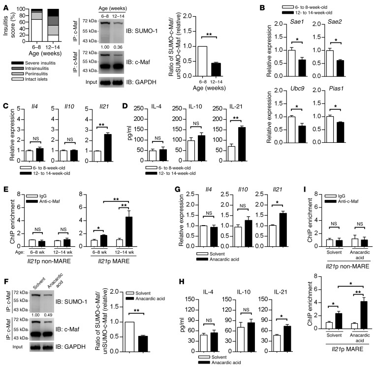
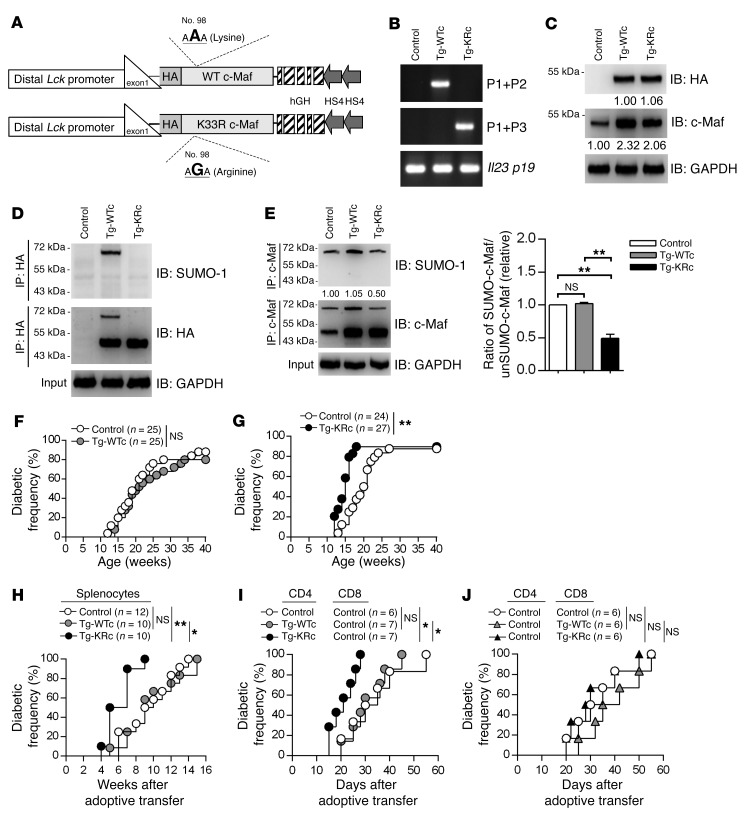
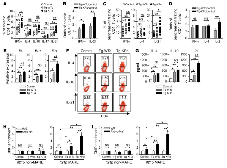
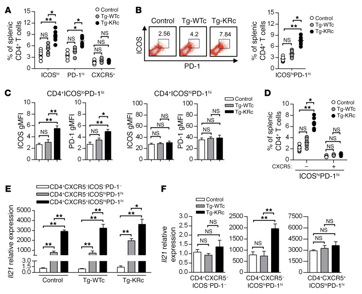
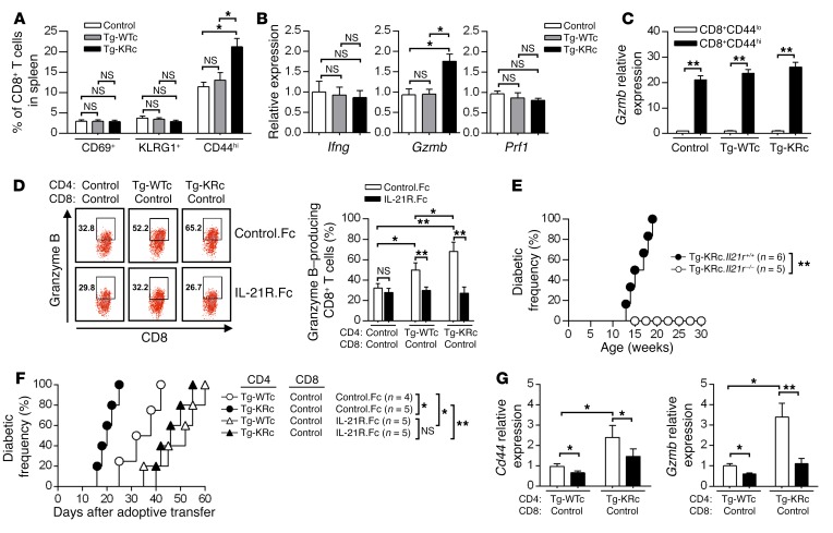
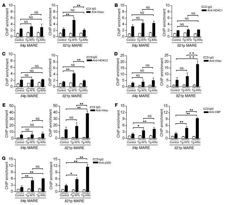
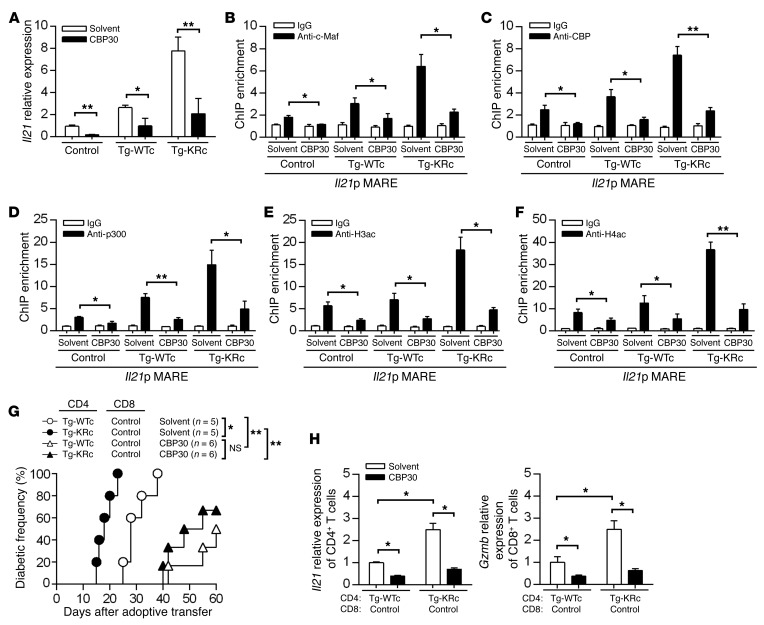
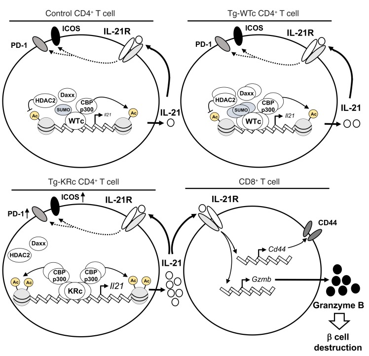
SUMOylation is involved in the development of several inflammatory diseases, but the physiological significance of SUMO-modulated c-Maf in autoimmune diabetes is not completely understood. Here, we report that an age-dependent attenuation of c-Maf SUMOylation in CD4+ T cells is positively correlated with the IL-21-mediated diabetogenesis in NOD mice. Using 2 strains of T cell-specific transgenic NOD mice overexpressing wild-type c-Maf (Tg-WTc) or SUMOylation site-mutated c-Maf (Tg-KRc), we demonstrated that Tg-KRc mice developed diabetes more rapidly than Tg-WTc mice in a CD4+ T cell-autonomous manner. Moreover, SUMO-defective c-Maf preferentially transactivated Il21 to promote the development of CD4+ T cells with an extrafollicular helper T cell phenotype and expand the numbers of granzyme B-producing effector/memory CD8+ T cells. Furthermore, SUMO-defective c-Maf selectively inhibited recruitment of Daxx/HDAC2 to the Il21 promoter and enhanced histone acetylation mediated by CREB-binding protein (CBP) and p300. Using pharmacological interference with CBP/p300, we illustrated that CBP30 treatment ameliorated c-Maf-mediated/IL-21-based diabetogenesis. Taken together, our results show that the SUMOylation status of c-Maf has a stronger regulatory effect on IL-21 than the level of c-Maf expression, through an epigenetic mechanism. These findings provide new insights into how SUMOylation modulates the pathogenesis of autoimmune diabetes in a T cell-restricted manner and on the basis of a single transcription factor.

Keywords: Autoimmunity; Cytokines; Epigenetics; Immunology; T cells.


Conflict of interest statement

Conflict of interest: The authors have declared that no conflict of interest exists.

Figures

Figure 1
Figure 1. c-Maf SUMOylation in CD4+ T cells is inversely correlated with the severity of insulitis and IL-21 production in NOD mice.
(A) The severity of insulitis was classified and scored on 100 islets from 10 NOD mice per group. Immunoprecipitation analysis of c-Maf SUMOylation in 6- to 8-week-old and 12- to 14-week-old NOD CD4+ cells cultured with anti-CD3 and anti-CD28 for 36 hours. (B and C) Expressions of Sae1, Sae2, Ubc9, and Pias1 mRNA (B) or Il4, Il10, and Il21 mRNA (C) in CD4+ cells cultured for 36 hours as described in A. (D) ELISA of indicated cytokines in supernatants of CD4+ T cells cultured as described in A for 48 hours. (E) ChIP analysis of the interaction of c-Maf with the Il21 promoter (Il21p) in CD4+ cells cultured for 36 hours as described in A. (F) Immunoprecipitation analysis of c-Maf SUMOylation in 6- to 8-week-old NOD CD4+ cells cultured for 36 hours with anti-CD3 and anti-CD28 in the presence of anacardic acid (3 μM) or its solvent (DMSO), which were added after 18 hours of culture. (G) Expressions of indicated cytokine mRNA in CD4+ T cells cultured for 36 hours as described in F. (H) ELISA of indicated cytokines in supernatants of CD4+ cells cultured as described in F for 48 hours. (I) ChIP analysis of the interaction of c-Maf with the Il21 promoter in CD4+ cells cultured for 36 hours as described in F. For E and I, isotype-matched IgG was used as a control. See complete unedited blots in the supplemental material. Data represent the mean ± SEM; n = 5 mice (A and F), n = 3 mice (BE and GI) per group; 3 independent experiments. *P < 0.05; **P < 0.01; 2-tailed Student’s t test (AD and FH) or 1-way ANOVA with Tukey’s post-test (E and I).
Figure 2
Figure 2. Accelerated kinetics of autoimmune diabetes in SUMO-defective c-Maf–transgenic NOD mice.
(A) Schematic diagram of distal Lck promoter–driven wild-type c-Maf or K33R c-Maf transgenes. (B) PCR detection of transgenic c-Maf in genomic DNA extracted from control, Tg-WTc, and Tg-KRc mice. Il23 p19 was used as an internal control. (C) Western blot analysis of c-Maf in control, Tg-WTc, and Tg-KRc CD4+ T cells. (D and E) Immunoprecipitation analysis of c-Maf SUMOylation in control, Tg-WTc, and Tg-KRc CD4+ T cells cultured with anti-CD3 and anti-CD28 for 36 hours. (F) Diabetes incidence in Tg-WTc NOD mice and their littermate controls. (G) Diabetes incidence in Tg-KRc NOD mice and their littermate controls. (H) Diabetes incidence in NOD-SCID recipients of splenocytes from 12- to 14-week-old control, Tg-WTc, or Tg-KRc NOD mice. (I and J) CD25CD4+ plus CD8+ T cells from 12- to 14-week-old indicated NOD mice were transferred into NOD.Rag1–/– mice; all groups were treated simultaneously in 2 independent experiments. The control CD4/control CD8 (white circles) group is presented in both I and J. Diabetic incidences among groups that received control CD8+ T cells plus different CD4+ T cells (I) and diabetic incidences in among groups that received control CD4+ T cells plus different CD8+ T cells (J) were compared with each other by a log-rank test. See complete unedited blots in the supplemental material. For E, data represent the mean ± SEM; n = 3 mice (B and C) or n = 5 mice (D and E) per group; 3 independent experiments (BE) or 2 independent experiments (H). *P < 0.05; **P < 0.01; 1-way ANOVA with Tukey’s post-test (E) or log-rank test (FJ).
Figure 3
Figure 3. Overexpression of SUMO-defective c-Maf preferentially augments IL-21–producing CD4+ T cells by enhancing its recruitment to the Il21p.
(A and B) Frequencies of IFN-γ+, IL-4+, IL-10+, IL-17+, and IL-21+ cells (A) and Tg/control ratio of IFN-γ+, IL-4+, and IL-21+ cells (B) in splenic CD4+ T cells from 12- to 14-week-old control, Tg-WTc, and Tg-KRc NOD mice were analyzed by flow cytometry. (C and D) Frequencies (C) and Tg/control ratio (D) of IFN-γ+, IL-4+, and IL-21+ cells in pancreas-infiltrating CD4+ T cells from 12- to 14-week-old control, Tg-WTc, and Tg-KRc NOD mice were analyzed by flow cytometry. (E) Expressions of Il4, Il10, and Il21 mRNA in naive control, Tg-WTc, and Tg-KRc NOD CD4+ T cells cultured with anti-CD3 and anti-CD28 for 36 hours. (F) Intracellular staining for IL-4, IL-10, and IL-21 in naive CD4+ T cells cultured as described in E for 48 hours. Numbers in outlined areas indicate the percentages of the gated populations. (G) ELISA of IL-4, IL-10, and IL-21 in supernatants of naive CD4+ T cells cultured as described in E for 48 hours. (H and I) ChIP analysis of the interaction of c-Maf with the Il21p in naive CD4+ T cells cultured for 36 hours as described in E. Isotype-matched IgG was used as a control. Data represent the mean ± SEM; n = 6 mice (A and B), n = 10 mice (C and D), n = 3 mice (EG), or n = 5 mice (H and I) per group; 3–4 independent experiments. *P < 0.05; **P < 0.01; 1-way ANOVA with Tukey’s post-test (A, C, E, and GI) or 2-tailed Student’s t test (B and D).
Figure 4
Figure 4. SUMO-defective c-Maf promotes the differentiation of CD4+ T cells with an extrafollicular helper T cell phenotype.
(A) Flow cytometry analysis of the expression of ICOS, PD-1, and CXCR5 in splenic CD4+ T cells from 12- to 14-week-old control, Tg-WTc, and Tg-KRc NOD mice. Summary of the frequencies of ICOShiCD4+, PD-1hiCD4+, and CXCR5+CD4+ T cells. (B) Flow cytometry analysis of the expression of ICOS and PD-1 in splenic CD4+ T cells as described in A. Numbers adjacent to outlined areas indicate the percentages of ICOShiPD-1hi cells (left panel). Right panel: Summary of the frequencies of ICOShiPD-1hiCD4+ T cells. (C) Flow cytometry analysis of ICOS or PD-1 expression in ICOSloPD-1loCD4+ and ICOShiPD-1hiCD4+ T cells from 12- to 14-week-old control, Tg-WTc, and Tg-KRc NOD mice. Summary of the geometric mean fluorescence intensity (gMFI) of ICOS or PD-1 in ICOSloPD-1loCD4+ or ICOShiPD-1hiCD4+ T cells. (D) Flow cytometry analysis of the expression of ICOS, PD-1, and CXCR5 in splenic CD4+ T cells as described in A. Summary of the frequencies of CXCR5ICOShiPD-1hiCD4+ and CXCR5+ICOShiPD-1hiCD4+ T cells. (E and F) Expression of Il21 mRNA in CXCR5ICOSloPD-1loCD4+, CXCR5ICOShiPD-1hiCD4+, and CXCR5+ICOShiPD-1hiCD4+ T cells from control, Tg-WTc, and Tg-KRc NOD mice. Data represent the mean ± SEM; n = 6 mice (AD) or n = 3 mice (E and F) per group; 3–4 independent experiments. *P < 0.05; **P < 0.01; 1-way ANOVA with Tukey’s post-test.
Figure 5
Figure 5. SUMO-defective c-Maf expands effector/memory CD8+ T cells and enhances their granzyme B production and diabetogenic activity in an IL-21–dependent manner.
(A) Flow cytometry analysis of CD69, KLRG1, and CD44 in splenic CD8+ T cells from 12- to 14-week-old control, Tg-WTc, and Tg-KRc NOD mice. Summary of the frequencies of CD69+CD8+, KLRG1+CD8+, and CD44hiCD8+ T cells. (B) Expression of Ifng, Gzmb, and Prf1 mRNA in splenic CD8+ T cells from 12- to 14-week-old control, Tg-WTc, and Tg-KRc NOD mice. (C) Expression of Gzmb mRNA in CD44loCD8+ and CD44hiCD8+ T cells from 12- to 14-week-old control, Tg-WTc, and Tg-KRc NOD mice. (D) Intracellular staining for granzyme B in control CD8+ T cells cocultured with control, Tg-WTc, and Tg-KRc CD4+ cells in Transwell chambers for 48 hours with anti-CD3 and anti-CD28 in the presence of control.Fc and IL-21R.Fc (2 μg/ml). Numbers adjacent to outlined areas indicate the percentages of granzyme B+ cells. Summary of the frequency of granzyme B+CD8+ T cells. (E) Diabetes incidence in Tg-KRc.Il21r+/+ and Tg-KRc.Il21r–/– NOD mice. (F) Diabetes incidence in NOD.Rag1–/– recipients injected with Tg-WTc and Tg-KRc CD25CD4+ T cells plus control CD8+ T cells on day 0, and then injected with control.Fc or IL-21R.Fc (10 μg) every 2 days from day 1 to day 13. (G) On day 14, quantitative reverse transcription PCR (RT-qPCR) analysis of Cd44 or Gzmb mRNA expression in CD8+ T cells from NOD.Rag1−/− recipients reconstituted as described in F. Data represent the mean ± SEM; n = 6 mice (A), n = 4 mice (BD), n = 3 mice (G) per group; 3 independent experiments (AD) or 2 independent experiments (F and G). *P < 0.05; **P < 0.01; 1-way ANOVA with Tukey’s post-test (A, B, D, and G), 2-tailed Student’s t test (C), or log-rank test (E and F).
Figure 6
Figure 6. SUMO-defective c-Maf prevents Daxx/HDAC2 recruitment to the Il21p and enhances CBP/p300-mediated histone acetylation.
(AC) ChIP analysis of the interaction of Daxx (A), HDAC1 (B), and HDAC2 (C) with the c-Maf–binding sites of the Il4p and the Il21p in naive control, Tg-WTc, and Tg-KRc CD4+ T cells cultured with anti-CD3 and anti-CD28 for 36 hours. (D and E) ChIP analysis of the abundance of H3ac (D) and H4ac (E) in the c-Maf–binding site of the Il4p or the Il21p in naive CD4+ T cells cultured for 36 hours as in AC. (F and G) ChIP analysis of the interaction of CBP (F) and p300 (G) with the c-Maf–binding site of the Il4p or the Il21p in naive CD4+ T cells cultured for 36 hours as in AC. Isotype-matched IgG was used as a control. Data represent the mean ± SEM; n = 3–5 mice per group; 3 independent experiments. *P < 0.05; **P < 0.01; 1-way ANOVA with Tukey’s post-test.
Figure 7
Figure 7. A CBP/p300 inhibitor attenuates SUMO-defective c-Maf–mediated transactivation of Il21 and ameliorates autoimmune diabetes in NOD mice.
(A) Expression of Il21 mRNA in naive control, Tg-WTc, and Tg-KRc CD4+ T cells cultured for 36 hours with anti-CD3 and anti-CD28 in the presence of CBP30 (2 μM) or its solvent (DMF), which were added after 18 hours of culture. (BD) ChIP analysis of the interaction of c-Maf (B), CBP (C), and p300 (D) with the c-Maf–binding site in the Il21p in naive CD4+ T cells cultured for 36 hours as in A. (E and F) ChIP analysis of the abundance of H3ac (E) and H4ac (F) in the c-Maf–binding site of the Il21p in naive CD4+ T cells cultured for 36 hours as in A. Isotype-matched IgG was used as a control. (G) Diabetic incidence in NOD.Rag1–/– recipients injected with effector Tg-WTc and Tg-KRc CD4+ T cells (CD4+CD25) plus control CD8+ T cells on day 0, and then injected with 2 mg/kg CBP30 or its solvent (DMF) every 2 days from day 1 to day 13. (H) On day 14, RT-qPCR analysis of Il21 mRNA expression in CD4+ T cells and Gzmb mRNA expression in CD8+ T cells from NOD.Rag1–/– recipients reconstituted as in G. Data represent the mean ± SEM; n = 3 mice (A and H), n = 5 mice (BF), n = 5–6 mice (G) per group; 3 independent experiments (AF) or 2 independent experiments (G and H). *P < 0.05; **P < 0.01; 2-tailed Student’s t test (AF), log-rank test (G), or 1-way ANOVA with Tukey’s post-test (H).
Figure 8
Figure 8. Schematic diagrams illustrating the critical role of SUMOylation in the regulation of c-Maf–mediated IL-21 production.

References

    1. Johnson ES. Protein modification by SUMO. Annu Rev Biochem. 2004;73:355–382. doi: 10.1146/annurev.biochem.73.011303.074118. - DOI - PubMed
    1. Liu B, Tahk S, Yee KM, Fan G, Shuai K. The ligase PIAS1 restricts natural regulatory T cell differentiation by epigenetic repression. Science. 2010;330(6003):521–525. doi: 10.1126/science.1193787. - DOI - PMC - PubMed
    1. Decque A, et al. Sumoylation coordinates the repression of inflammatory and anti-viral gene-expression programs during innate sensing. Nat Immunol. 2016;17(2):140–149. - PubMed
    1. Ding X, et al. Protein SUMOylation is required for regulatory T cell expansion and function. Cell Rep. 2016;16(4):1055–1066. doi: 10.1016/j.celrep.2016.06.056. - DOI - PubMed
    1. Geiss-Friedlander R, Melchior F. Concepts in sumoylation: a decade on. Nat Rev Mol Cell Biol. 2007;8(12):947–956. doi: 10.1038/nrm2293. - DOI - PubMed

Publication types

MeSH terms