. 2018 Sep 15:78:165-177.
doi: 10.1016/j.actbio.2018.07.048. Epub 2018 Jul 29.

Glial-derived growth factor and pleiotrophin synergistically promote axonal regeneration in critical nerve injuries

Affiliations

Glial-derived growth factor and pleiotrophin synergistically promote axonal regeneration in critical nerve injuries

Nesreen Zoghoul Alsmadi et al. Acta Biomater. .

Abstract

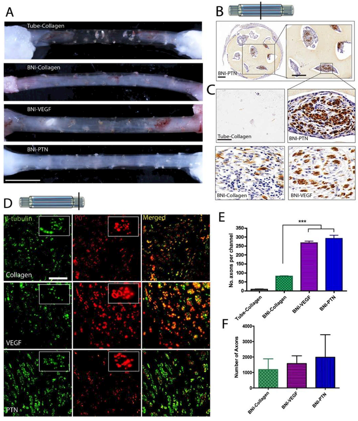
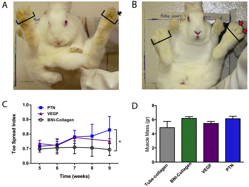
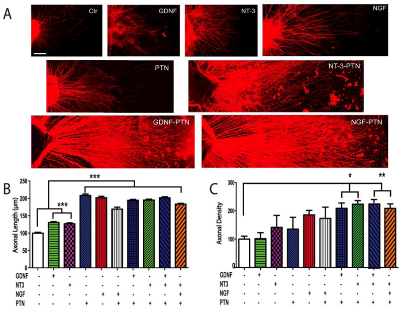
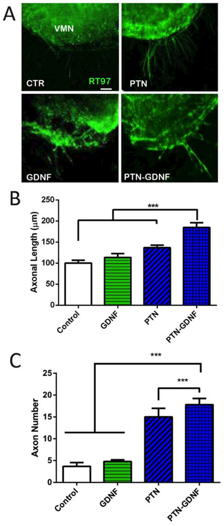
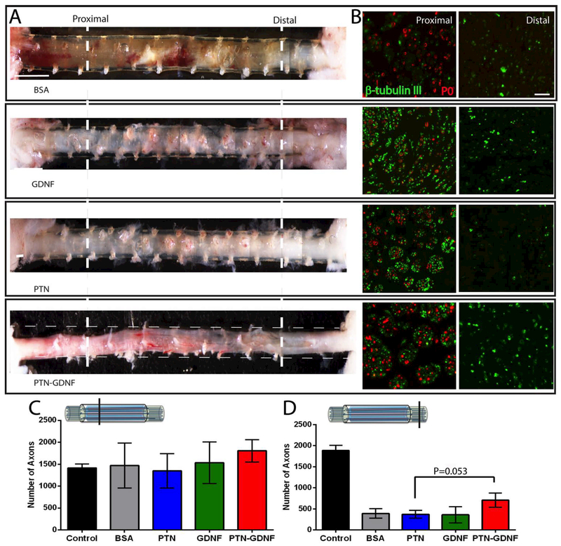
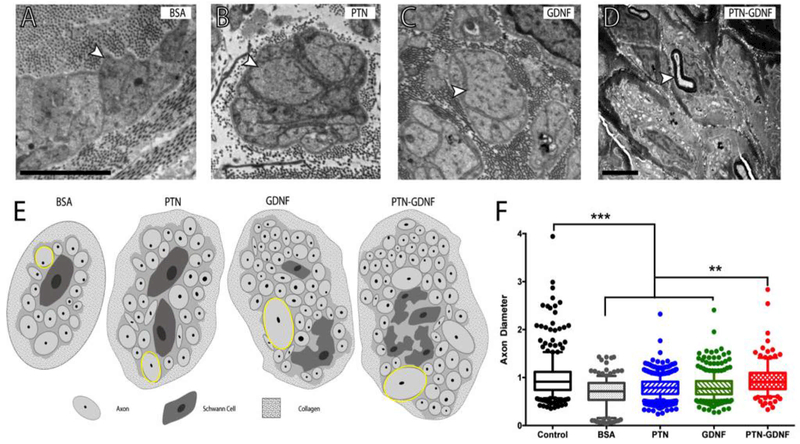
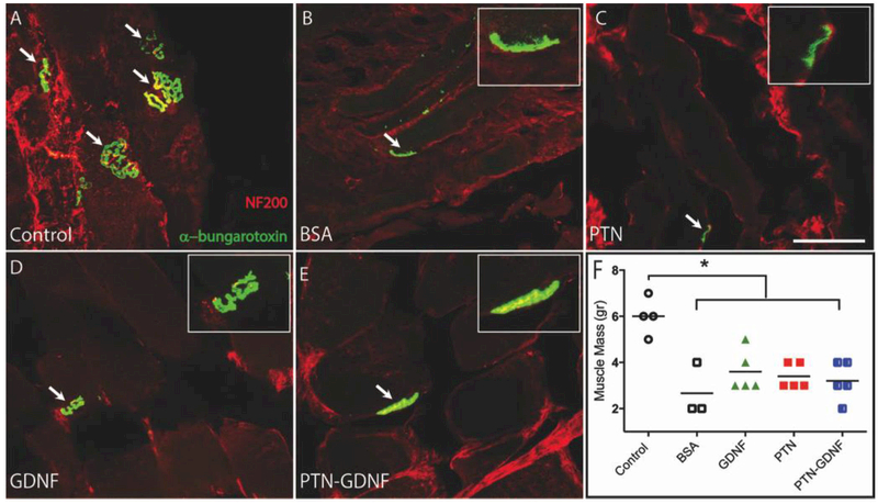
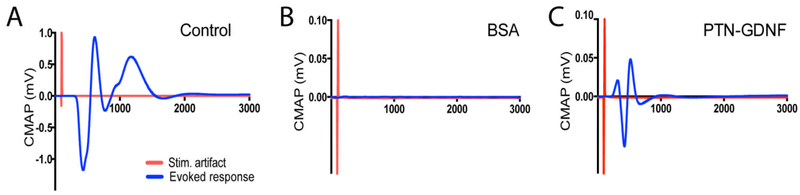
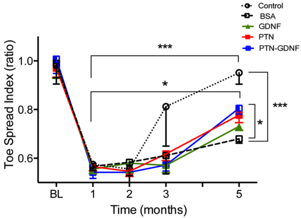
The repair of nerve gap injuries longer than 3 cm is limited by the need to sacrifice donor tissue and the morbidity associated with the autograft gold standard, while decellularized grafts and biodegradable conduits are effective only in short nerve defects. The advantage of isogenic nerve implants seems to be the release of various growth factors by the denervated Schwann cells. We evaluated the effect of vascular endothelial growth factor, neurotrophins, and pleiotrophin (PTN) supplementation of multi-luminal conduits, in the repair of 3 and 4 cm nerve gaps in the rabbit peroneal nerve. In vitro screening revealed a synergistic regenerative effect of PTN with glial-derived neurotrophic factor (GDNF) in promoting sensory axon density, and motor axonal growth from spinal cord explants. In vivo, pleiotrophins were able to support nerve regrowth across a 3 cm gap. In the 4 cm lesions, PTN-GDNF had a modest effect in the number of axons distal to the implant, while increasing the mean axon diameter (1 ± 0.4; p ≤ 0.001) over PTN or GDNF alone (0.80 ± 0.2, 0.84 ± 0.5; respectively). Some regenerated axons reinnervated muscle targets as indicated by neuromuscular junction staining. However, many were wrapped in Remak bundles, suggesting a delay in axonal sorting, explaining the limited electrophysiological function of the reinnervated muscle, and the modest recovery in toe spreading in the PTN-GDNF repaired animals. These results support the use of synergistic neurotrophic/pleiotrophic growth factors in long gap repair and underscore the need for re-myelination strategies distal to the injury site.

Statement of significance: Nerve injuries due to trauma or tumor resection often result in long gaps that are challenging to repair. The best clinical option demands the use of autologous grafts that are associated with serious side effects. Bioengineered nerves are considered a good alternative, particularly if supplemented with growth factors, but current options do not match the regenerative capacity of autografts. This study revealed the synergistic effect of neurotrophins and pleiotrophins designed to achieve a broad cellular regenerative effect, and that GDNF-PTN are able to mediated axonal growth and partial functional recovery in a 4 cm nerve gap injury, albeit delays in remyelination. This report underscores the need for defining an optimal growth factor support for biosynthetic nerve implants.

Keywords: Agarose; Long gap; Micro-channels; Neurotrophins; Pleiotrophins; Remak bundles.


Conflict of interest statement

Competing Interests

The authors declare that they have no competing interests.

Figures

Figure 1.
Figure 1.
Experimental design. The flow chart indicates the three phases of the study, the groups, the sample size and timelines for the repair studies.
Figure 2.
Figure 2.. Toe spread apparatus.
(A) Photograph of the TSI custom device shows a rabbit secured to a sliding hook via a harness on the top of the device. Removing of the metal element causes the rabbit to free fall and then come to a halt resulting in a startle response. (B) Baseline shows left, and right foot has equal toe spreads. (C) One month after injury shows a loss of toe spread in the injured foot (D) Five months after injury shows the recovery of the toe spread in the left foot.
Figure 3.
Figure 3.. BNI with multi-luminal growth factor release for nerve gap repair.
(A) Cross section schematic of the BNI with MPs mixed in collagen. (B) Release profile of PTN-MPs. Inserts: SEM images of the PTN-MPs and confocal images of DRG axonal growth (β-tubulin; red) demonstrating the bioactivity of the encapsulated growth factors. C) COMSOL model of PTN diffusion in the BNI microchannels overtime. (D) BNI fabrication: (i) placement of metal rods in the silicone conduit and filled with agarose, (ii) rod removal and MPs/collagen loading, (iii) implantable device. (E) Cross section of the BNI showing the eight microchannels with luminal MP-collagen (insert). (F) BNI sutured to both ends of the injured common peroneal (CP) nerve. Tib = tibial nerve. Scale bars: B) SEM: 5 μm, DRG: 1 mm, D) 1 mm, E) Device: 250 μm, Insert: 10 μm. F) 2 mm.
Figure 4.
Figure 4.. Effect of PTN and VEGF in nerve regeneration across a 3 cm-long nerve gap.
(A) Photographs of regenerated nerves 9 weeks after implantation. Collagen-filled conduits failed to mediate nerve growth. Conversely, BNIs with collagen, VEGF-MPs or PTN-MPs showed nerve regeneration. (B) Axonal growth in the BNI was confirmed by positive NFP staining. (C) Representative growth inside the microchannels at the middle of the conduit is shown at higher magnification. (D) Double labeling of axons (β-tubulin) and myelin (P0), confirmed nerve regeneration across the gap. (E) The number of axons per channel and (F) distal to the implant showed a mild effect of PTN. *** = p ≤ 0.001. Scale bars: A) 0.5 cm, (B) 350 μm, (C and D) 50 μm.
Figure 5.
Figure 5.. Motor functional recovery after a 3 cm gap repair with BNI-PTN.
(A) All injured animals were unable to show digit abduction (black arrow) up to 6 weeks after injury repair. (B, C) By week 9, those implanted with PTN MPs showed significant improvement in toe spreading (red arrows) compared to those with collagen filled conduits. (D) None of the groups showed a significant recovery in the total mass of the tibialis anterior muscle at this time. * = p ≤ 0.05.
Figure 6.
Figure 6.. Synergistic growth factor effects on sensory axons in vitro.
(A) Confocal images of neonatal DRG with neurotrophin/pleiotrophin support for axonal growth. (B) Quantitative analysis revealed that PTN, alone or in combination, doubled the axonal length, and that (C) the combination of PTN with GDNF or NT-3 increased the axonal density (number of regenerating axons) compared to control. Axonal density is reported as a percentage of the negative control. *** = p ≤ 0.001, ** = p ≤ 0.01, * = p ≤ 0.05. Scale bar 10 μm.
Figure 7.
Figure 7.. Enhanced motor nerve growth in SC slices by PTN-GDNF.
(A) Axon growth from the ventral SC explants. (B) PTN-GDNF showed a significant improvement on axon length compared to control, or PTN and GDNF alone. (C) Compared to control and GDNF, PTN was able to show a significant effect in the number of extending axons, which was further improved in the PTN-GDNF group. The mean represents the average of 20 axons per SC slice and four separate experiments per group. ***=p ≤ 0.001. Scale Bar 50 μm
Figure 8.
Figure 8.. Effect of PTN-GDNF on nerve regeneration across a 4 cm nerve gap.
(A) Photographs of the regenerated nerves in the conduits or with the conduit indicated by longitudinal broken lines. (B) β-tubulin/P0 immunofluorescence at 1 cm (proximal) and 3 cm (distal) from the proximal end. Enhanced axonal regeneration is evidenced in the PTN-GDNF group. The number of axons proximally (C) is comparable among the groups, but (D) reduced distally in those with BNI implants, where PTN-GDNF showed a trend towards significance over the PTN group (p=0.053). Scale Bar: A) 600 μm, B) 50 μm.
Figure 9.
Figure 9.. Radial sorting delay.
Electron micrographs of axons in Remak bundles. (A) Compared to those with BSA, larger axons were observed in the (B) PTN and (C) GDNF implants (arrowheads). (D) Re-myelinated axons were only present in the PTN-GDNF group. (E) Schematic representation of axons in the Remak bundles per treatment. (F) Axon diameter distribution showed significantly smaller axons in the BSA, PTN and GDNF groups compared to the PTN-GDNF. Scale bar: 5 μm. *** = p ≤ 0.001, ** = p ≤ 0.01.
Figure 10.
Figure 10.. Re-innervation of the tibialis anterior muscle.
(A) Co-labeling of axons (NF200) and AchR clustering (α-bungarotoxin) demonstrated the successful target re-innervation. The amount of axon/NMJs overlap was larger in the GDNF (D) and PTN-GDNF (E) compared to the BSA (B) and PTN (C) groups, but not as elaborate as the (cut-resuture) control. (F) Muscle mass did not improve significantly compared to the positive control. Scale bar: 5 μm. * = p ≤ 0.05.
Figure 11.
Figure 11.. Evoked compound action potentials from regenerated nerves.
(A) Compound action potentials were evoked in the cut-resuture control but failed in all animals implanted with (B) BSA, GDNF or PTN BNIs. (C) Only 1 of 5 rabbits repaired with PTN-GDNF showed a CMAP with an approximately 10% of the amplitude generated in the control group.
Figure 12
Figure 12. PTN-GDNF mediates moderate functional recovery after critical nerve gap repair.
Evaluation of toe-spread index after five months confirmed that animals implanted with PTN-GDNF BNIs were significantly better compared to those implanted with BSA, but not compared to the positive control. *** = p ≤ 0.001, * = p ≤ 0.05.

References

    1. Höke A, Brushart T, Introduction to special issue: Challenges and opportunities for regeneration in the peripheral nervous system, Exp. Neurol 223 (2010) 1–4. doi:10.1016/j.expneurol.2009.12.001. - DOI - PMC - PubMed
    1. Grinsell D, Keating CP, Peripheral Nerve Reconstruction after Injury: A Review of Clinical and Experimental Therapies, Biomed Res. Int 2014 (2014). doi:10.1155/2014/698256. - DOI - PMC - PubMed
    1. Fu SY, Gordon T, Contributing factors to poor functional recovery after delayed nerve repair: prolonged denervation., J. Neurosci 15 (1995) 3886–3895. - PMC - PubMed
    1. Krarup C, Archibald SJ, Madison RD, Factors that influence peripheral nerve regeneration: An electrophysiological study of the monkey median nerve, Ann. Neurol 51 (2002) 69–81. doi:10.1002/ana.10054. - DOI - PubMed
    1. Moore AM, Kasukurthi R, Magill CK, Farhadi FH, Borschel GH, Mackinnon SE, Limitations of conduits in peripheral nerve repairs, Hand 4 (2009) 180–186. doi:10.1007/s11552-008-9158-3. - DOI - PMC - PubMed

Publication types

MeSH terms