. 2018 Sep 1;159(9):3365-3377.
doi: 10.1210/en.2018-00531.

Sex-Dimorphic and Sex Hormone-Dependent Role of Steroid Sulfatase in Adipose Inflammation and Energy Homeostasis

Affiliations

Sex-Dimorphic and Sex Hormone-Dependent Role of Steroid Sulfatase in Adipose Inflammation and Energy Homeostasis

Yuhan Bi et al. Endocrinology. .

Abstract

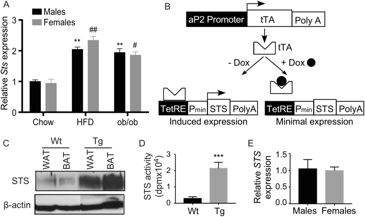
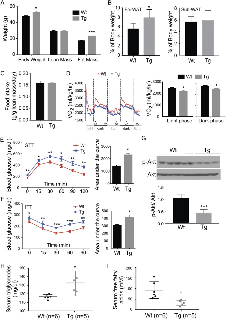
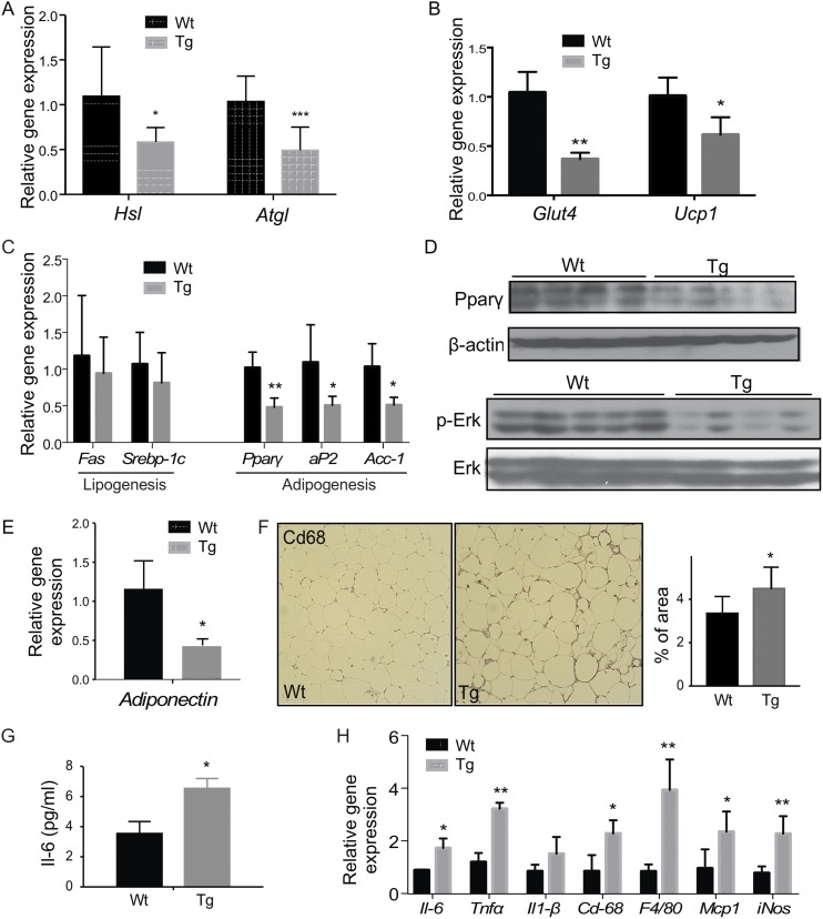
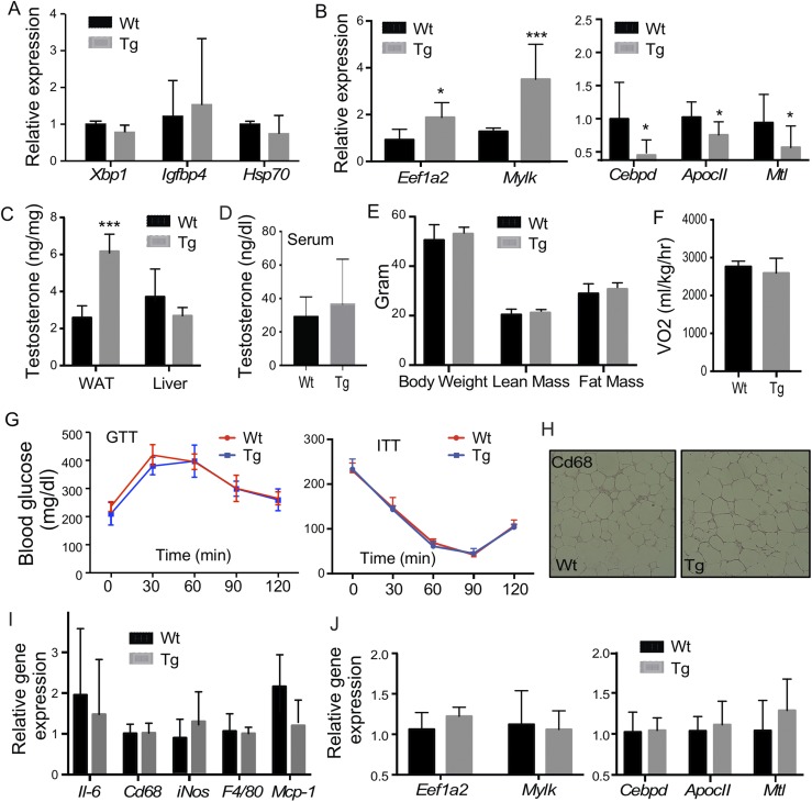
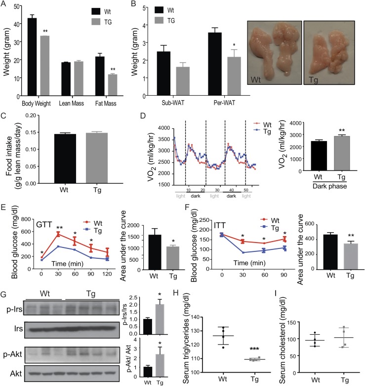
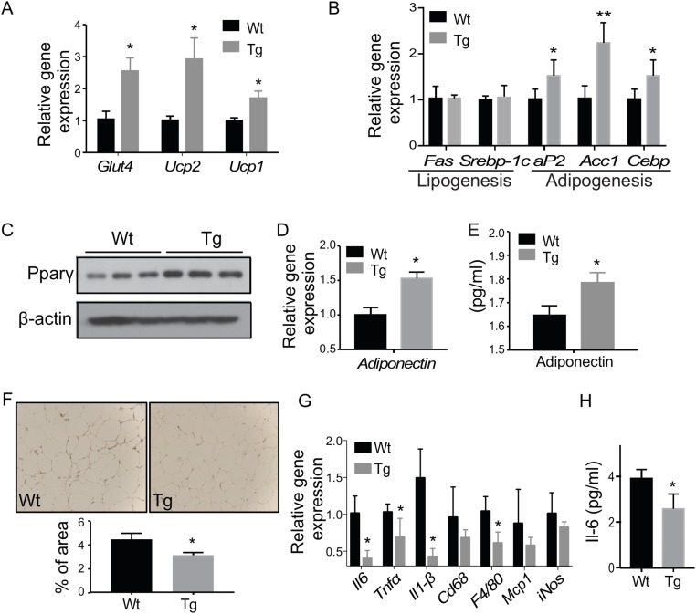
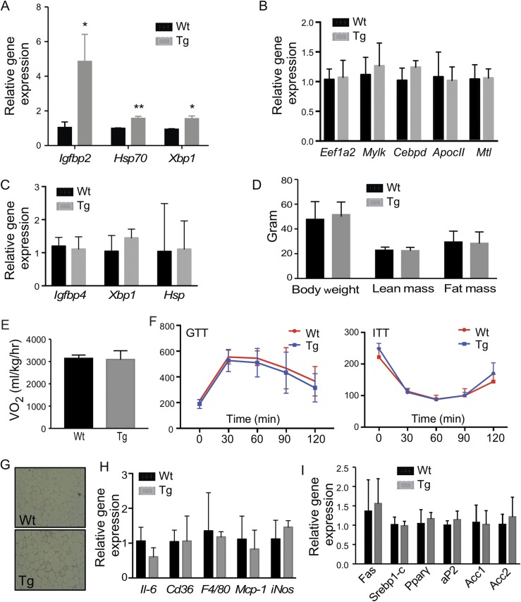
Steroid sulfatase (STS), a desulfating enzyme that converts steroid sulfates to hormonally active steroids, plays an important role in the homeostasis of sex hormones. STS is expressed in the adipose tissue of both male and female mice, but the role of STS in the development and function of adipose tissue remains largely unknown. In this report, we show that the adipose expression of Sts was induced in the high-fat diet (HFD) and ob/ob models of obesity and type 2 diabetes. Transgenic overexpression of the human STS in the adipose tissue of male mice exacerbated the HFD-induced metabolic phenotypes, including increased body weight gain and fat mass, and worsened insulin sensitivity, glucose tolerance, and energy expenditure, which were accounted for by adipocyte hypertrophy, increased adipose inflammation, and dysregulation of adipogenesis. The metabolic harm of the STS transgene appeared to have resulted from increased androgen activity in the adipose tissue, and castration abolished most of the phenotypes. Interestingly, the transgenic effects were sex specific, because the HFD-fed female STS transgenic mice exhibited improved metabolic functions, which were associated with attenuated adipose inflammation. The metabolic benefit of the STS transgene in female mice was accounted for by increased estrogenic activity in the adipose tissue, whereas such benefit was abolished upon ovariectomy. Our results revealed an essential role of the adipose STS in energy homeostasis in sex- and sex hormone-dependent manner. The adipose STS may represent a therapeutic target for the management of obesity and type 2 diabetes.


Figures

Figure 1.
Figure 1.
Adipose induction of STS in obese mice, and creation of Tg mice expressing STS in the adipose tissue. (A) The mRNA expression of mouse Sts in the epididymal WAT of chow-fed wild-type (Wt) mice, HFD-fed Wt mice, and chow-fed ob/ob mice. (B) The schematic representation of the Tet-off STS Tg system. (C) The expression of STS protein in the epi-WAT and brown adipose tissue (BAT) of the Wt and Tg mice was measured by Western blotting. (D) The adipose STS enzymatic activity was determined by estrone sulfate conversion assay and was normalized against protein concentrations. (E) The adipose expression of the STS transgene in female and male Tg mice was measured by real-time PCR. n = 4 mice per group. * or #, P < 0.05; ** or ##, P < 0.01; ***P < 0.001, compared with (A) chow of the sex, or (D) or compared with the Wt. Pmin, minimal human cytomegalovirus promoter; PolyA, polyacrylamide; SV40 polyA, simian virus 40 polyadenylation signal.
Figure 2.
Figure 2.
Adipose overexpression of STS aggravates HFD-induced adiposity, insulin resistance, and glucose intolerance in male mice. All mice are males. Mice were fed with HFD for 20 weeks before analysis. Mice were analyzed for (A) body weight and body composition, (B) fat mass, (C) food intake, (D) oxygen consumption, (E) GTT, and (F) ITT. The quantifications of the GTT and ITT results are shown as the areas under the curve. (G) Western blot analysis of Akt phosphorylation in epi-WAT. Shown below is the densitometric quantification of the Western blotting results. The serum levels of (H) triglycerides and (I) free fatty acids. Results are expressed as mean ± SD. n = 4 mice per group, except those labeled in (H) and (I). *P < 0.05; **P < 0.01, compared with the Wt.
Figure 3.
Figure 3.
Adipose overexpression of STS decreases lipolysis and adipogenesis and aggravates HFD-induced adipose and systemic inflammation in males. Mice are the same as described in Fig. 2. The epi-WAT expression of genes responsible for (A) lipolysis, (B) glucose uptake and energy expenditure, and (C) lipogenesis and adipogenesis was measured by real-time PCR. (D) The protein level of PPARγ and ERK1/2 was measured by Western blotting. (E) Epi-WAT expression of adiponectin. (F) Immunostaining of Cd68. Shown on the right is the quantification. The (G) serum level of IL-6 and (H) adipose expression on proinflammatory genes and macrophage marker genes. Results are expressed as mean ± SD; n = 4 mice per group. *P < 0.05; **P < 0.01; ***P < 0.005, compared with the Wt.
Figure 4.
Figure 4.
The metabolic phenotype in aP2-STS Tg males is androgen dependent. Mice are the same as described in Fig. 2. Shown are (A) adipose expression of estrogen-responsive genes, (B) adipose expression of androgen-responsive genes, (C) adipose and liver levels of testosterone, and (D) the serum levels of testosterone. Male mice were castrated before being fed with HFD for 20 weeks. Shown are (E) body weight and body composition, (F) oxygen consumption, (G) GTT and ITT, (H) immunostaining of CD68, (I) adipose expression of proinflammatory genes and macrophage marker genes, and (J) adipose expression of androgen responsive genes. Results are expressed as mean ± SD; n = 4 mice per group for all panels, except (B) and (C): Wt, n = 6; Tg, n = 5. *P < 0.05; **P < 0.01; ***P < 0.005, compared with the Wt.
Figure 5.
Figure 5.
Adipose overexpression of STS improves metabolic functions in HFD-fed females. All mice are females. Mice were fed with HFD for 20 weeks before analysis. Mice were analyzed for (A) body weight and body composition, (B) fat mass, (C) food intake, (D) oxygen consumption, (E) GTT, and (F) ITT. The quantifications of the GTT and ITT results are shown as the areas under the curve. (G) Western blot analysis of Irs-1 and Akt phosphorylation in epi-WAT. Shown on the right are the densitometric quantifications of the Western blotting results. The serum levels of (H) triglycerides and (I) cholesterol. Results are expressed as mean ± SD; n = 4 mice per group. *P < 0.05; **P < 0.01, compared with the Wt. sub-WAT, subcutaneous WAT.
Figure 6.
Figure 6.
Adipose overexpression of STS increases energy expenditure and adipogenesis and ameliorates HFD-induced adipose and systemic inflammation in females. Mice are the same as described in Fig. 5. (A) Adipose expression of genes responsible for energy uptake and expenditure, and (B) lipogenesis and adipogenesis was measured by real-time PCR. (C) The protein level of PPARγ was measured by Western blotting. The (D) adipose mRNA expression of adiponectin and (E) the serum level of adiponectin. (F) Immunostaining of Cd68. Shown below is the quantification. Adipose expression of (G) proinflammatory genes and macrophage marker genes and (H) the serum level of IL-6. Results are expressed as mean ± SD; n = 4 mice per group. *P < 0.05; **P < 0.01, compared with the Wt.
Figure 7.
Figure 7.
The metabolic benefit in female Tg mice is estrogen dependent. Mice are the same as described in Fig. 5. Shown are adipose expression of (A) estrogen-responsive genes and (B) androgen-responsive genes as measured by real-time PCR. Female mice were ovariectomized before being fed with HFD for 20 weeks. Shown are (C) adipose expression of estrogen-responsive genes, (D) body weight and body composition analysis, (E) oxygen consumption, (F) GTT and ITT, (G) immunostaining of CD68, (H) adipose expression of proinflammatory genes and macrophage marker genes, and (I) genes involved in adipogenesis and lipogenesis. Results are expressed as mean ± SD; n = 4 mice per group. *P < 0.05; **P < 0.01, compared with the Wt.

References

    1. Oh JY, Barrett-Connor E, Wedick NM, Wingard DL; Rancho Bernardo Study . Endogenous sex hormones and the development of type 2 diabetes in older men and women: the Rancho Bernardo study. Diabetes Care. 2002;25(1):55–60. - PubMed
    1. Navarro G, Allard C, Xu W, Mauvais-Jarvis F. The role of androgens in metabolism, obesity, and diabetes in males and females. Obesity (Silver Spring). 2015;23(4):713–719. - PMC - PubMed
    1. Carr MC. The emergence of the metabolic syndrome with menopause. J Clin Endocrinol Metab. 2003;88(6):2404–2411. - PubMed
    1. Smith EP, Boyd J, Frank GR, Takahashi H, Cohen RM, Specker B, Williams TC, Lubahn DB, Korach KS. Estrogen resistance caused by a mutation in the estrogen-receptor gene in a man. N Engl J Med. 1994;331(16):1056–1061. - PubMed
    1. Morishima A, Grumbach MM, Simpson ER, Fisher C, Qin K. Aromatase deficiency in male and female siblings caused by a novel mutation and the physiological role of estrogens. J Clin Endocrinol Metab. 1995;80(12):3689–3698. - PubMed

Publication types

MeSH terms