. 2018 Nov 2;46(19):10368-10379.
doi: 10.1093/nar/gky676.

Structural basis of chimpanzee APOBEC3H dimerization stabilized by double-stranded RNA

Affiliations

Structural basis of chimpanzee APOBEC3H dimerization stabilized by double-stranded RNA

Tatsuya Matsuoka et al. Nucleic Acids Res. .

Abstract

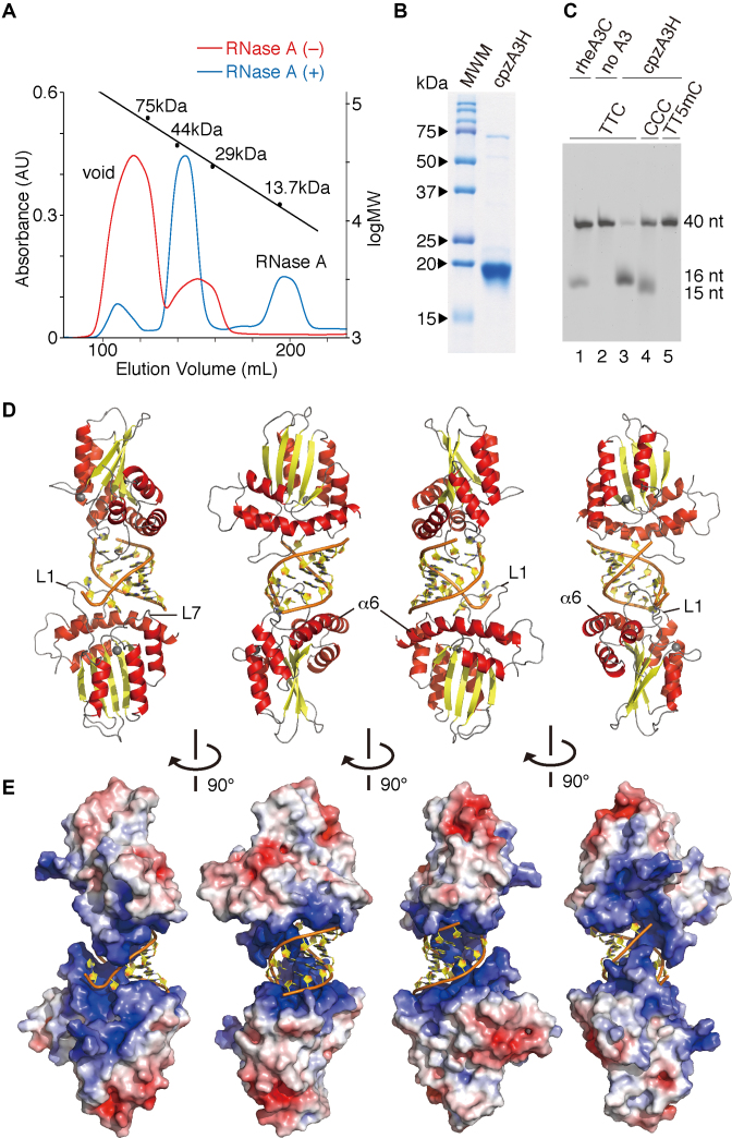
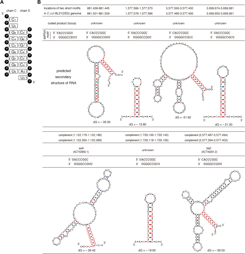
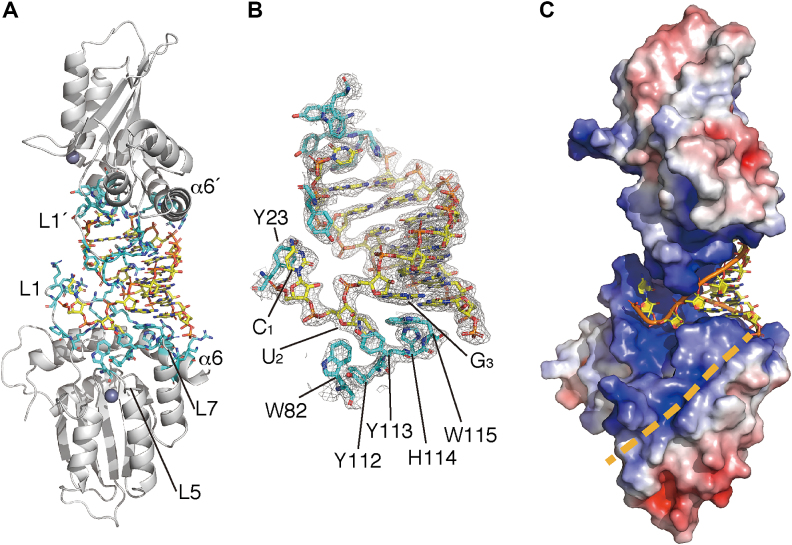
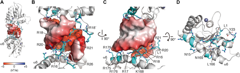
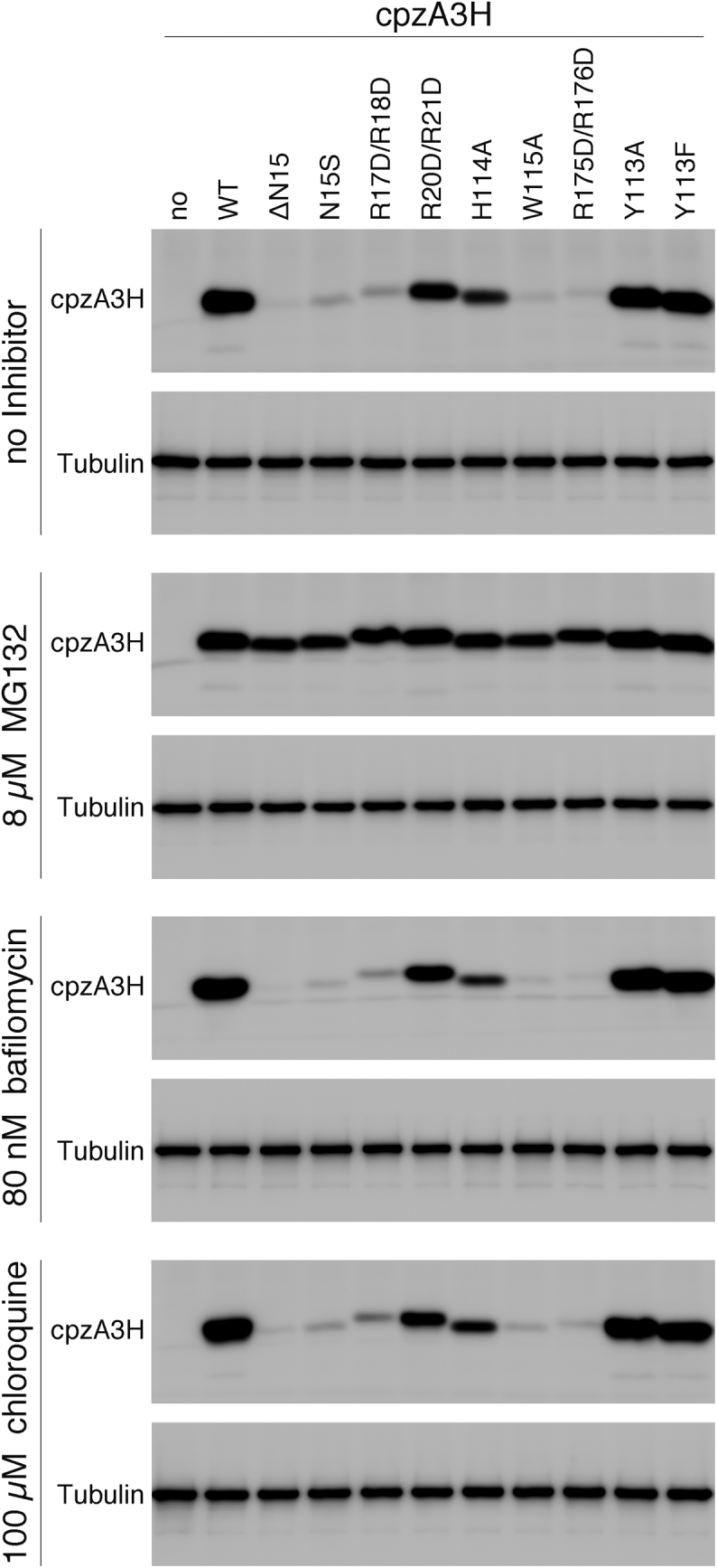
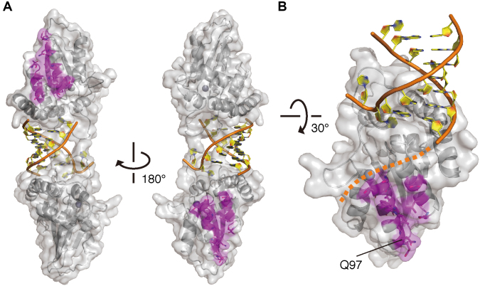
APOBEC3H (A3H) is a mammal-specific cytidine deaminase that potently restricts the replication of retroviruses. Primate A3Hs are known to exert key selective pressures against the cross-species transmission of primate immunodeficiency viruses from chimpanzees to humans. Despite recent advances, the molecular structures underlying the functional mechanisms of primate A3Hs have not been fully understood. Here, we reveal the 2.20-Å crystal structure of the chimpanzee A3H (cpzA3H) dimer bound to a short double-stranded RNA (dsRNA), which appears to be similar to two recently reported structures of pig-tailed macaque A3H and human A3H. In the structure, the dsRNA-binding interface forms a specialized architecture with unique features. The analysis of the dsRNA nucleotides in the cpzA3H complex revealed the GC-rich palindrome-like sequence preference for dsRNA interaction, which is largely determined by arginine residues in loop 1. In cells, alterations of the cpzA3H residues critical for the dsRNA interaction severely reduce intracellular protein stability due to proteasomal degradation. This suggests that cpzA3H stability is regulated by the dsRNA-mediated dimerization as well as by unknown cellular machinery through proteasomal degradation in cells. Taken together, these findings highlight unique structural features of primate A3Hs that are important to further understand their cellular functions and regulation.


Figures

Figure 1.
Figure 1.
Crystal structure of the cpzA3H dimer with an RNA duplex. (A) Gel filtration Superdex 75 elution profile of cpzA3H with (blue) or without (red) RNase A treatment. The standard MW marker proteins conalbumin (75 kDa), ovalbumin (44 kDa), carbonic anhydrase (29 kDa) and RNase A (13.7 kDa) were used. (B) Gel image of purified cpzA3H stained with Coomassie brilliant blue R-250; MWM, MW marker. (C) Deamination activity of cpzA3H. Forty-nucleotide ssDNA substrates containing TTC (lanes 1, 2 and 3), the CCC (lane 4) or the TT5mC (lane 5) motifs were used in the UDG-dependent deamination assay. The recombinant protein rhesus macaque A3C (rheA3C) (lane 1) was used as a control; 5mC, 5-methylcytosine. (D) Ribbon and (E) electrostatic surface representations of cpzA3H seen in four rotated views. The ribbons are colored according to the secondary structures: α-helices (red); β-strands (yellow); loops (gray) and dsRNA (orange/yellow). Coordinated zinc ions are represented as navy spheres. The loops 1 and 7 and the α6 helix in chain A are indicated as L1, L7 and α6, respectively. The accessible surface area of cpzA3H is colored according to the calculated electrostatic potential from –5.0 kT/e (red) to +5.0 kT/e (blue).
Figure 2.
Figure 2.
Identification of Escherichia coli-derived nucleotide sequences analogous to the dsRNA detected in the cpzA3H crystal structure. (A) Diagram of the dsRNA secondary structure modeled in the crystal structure. (B) Seven of our identified nucleotide sequences and their putative RNA secondary structures are shown. The dsRNA sequences identified in the crystal structure are shown as the ‘dsRNA motif’, consisting of 5′-YYRCCGGGU and 5′-YACCCGGY (Y, pyrimidine—C or U; R, purine—G or A). Using an ‘in-house’ program, the locations of these two nucleotide sequences were determined in the complete genome of E. coli BL21(DE3), which was used for cpzA3H protein expression. Based on the genome sequences around the identified positions, the RNA secondary structures were predicted with the mfold program. The nucleotides analogous to the dsRNA motif are highlighted in red. No dsRNA motifs were identified in the GST-cpzA3H expression plasmid.
Figure 3.
Figure 3.
Interactions between cpzA3H proteins and the RNA duplex. (A) Overall structure of cpzA3H–dsRNA complex. The cpzA3H and the dsRNA are represented with gray ribbons and orange/yellow sticks, respectively. cpzA3H residues responsible for the RNA interaction are highlighted with cyan sticks. The loop 5 is indicated as L5. L1′ and α6′ are in chain B. (B) The electron density map (2FO-FC, contoured at the 1.5σ level) around the termini of the RNA duplex. The aromatic residues involved in the nucleobase interactions are highlighted with cyan sticks. (C) A hypothetical path for the extended ssRNA portion (beige dashed line). The electrostatic surface potential is colored between –5.0 kT/e (red) and +5.0 kT/e (blue).
Figure 4.
Figure 4.
Roles of A3H loop 1 (L1) and the α6 helix (α6) in the interaction with dsRNA. (A) RNA–duplex interaction interfaces formed by positively charged cpzA3H residues. The overall complex structure is represented with gray ribbons (cpzA3H) and an electrostatic surface (RNA). The basic residues involved in the RNA interaction are represented with cyan sticks. (B) Enlarged view of the interaction site between L1 and dsRNA. Basic residues R17, R18, R20 and R21 of L1 protrude into the negatively charged major groove of dsRNA. (C) Basic residues of L1 and α6 are aligned along the curvature of the RNA phosphate backbone. (D) The α6 position is fixed via the formation of a hydrogen bond between the L1 stem N15 (gray stick) and the α6 L166. The structure is shown without dsRNA is represented.
Figure 5.
Figure 5.
Effect of cpzA3H mutations in the RNA recognition interface on the intracellular levels of cpzA3H. WT cpzA3H and mutants were transiently expressed in 293T cells in the absence of an inhibitor or in the presence of MG132 (8 μM), chloroquine (100 μM) or bafilomycin A1 (80 nM). The proteins were detected by western blotting with the anti-DYKDDDDK tag monoclonal antibody (Anti-FLAG) or anti-β-tubulin antibody (Anti-β-Tubulin).
Figure 6.
Figure 6.
A view of the Vif-interaction interface in the cpzA3H dimer. (A) Two separate A3H sites are responsible for Vif binding. cpzA3H and dsRNA are represented in gray ribbons with a transparent surface and orange/yellow ribbons with nucleobases, respectively. The residues (magenta sticks) critical for HIV-1–Vif interaction are mapped on the dimer structure. (B) Highlighted view of the Vif-binding interface and its unique residue Q97 in the chimpanzee-bonobo lineage. Residue Q97 is one of the most important determinants for the differential sensitivity of HIV-1/SIVcpz Vifs. A putative path for the extended ssRNA portion is also shown (orange dashed line).

References

    1. Desimmie B.A., Delviks-Frankenberrry K.A., Burdick R.C., Qi D., Izumi T., Pathak V.K.. Multiple APOBEC3 restriction factors for HIV-1 and one Vif to rule them all. J. Mol. Biol. 2014; 426:1220–1245. - PMC - PubMed
    1. Harris R.S., Dudley J.P.. APOBECs and virus restriction. Virology. 2015; 479–480:131–145. - PMC - PubMed
    1. Iwatani Y., Chan D.S., Wang F., Maynard K.S., Sugiura W., Gronenborn A.M., Rouzina I., Williams M.C., Musier-Forsyth K., Levin J.G.. Deaminase-independent inhibition of HIV-1 reverse transcription by APOBEC3G. Nucleic Acids Res. 2007; 35:7096–7108. - PMC - PubMed
    1. Chaurasiya K.R., McCauley M.J., Wang W., Qualley D.F., Wu T., Kitamura S., Geertsema H., Chan D.S., Hertz A., Iwatani Y. et al. . Oligomerization transforms human APOBEC3G from an efficient enzyme to a slowly dissociating nucleic acid-binding protein. Nat. Chem. 2014; 6:28–33. - PMC - PubMed
    1. Newman E.N., Holmes R.K., Craig H.M., Klein K.C., Lingappa J.R., Malim M.H., Sheehy A.M.. Antiviral function of APOBEC3G can be dissociated from cytidine deaminase activity. Curr. Biol. 2005; 15:166–170. - PubMed

Publication types