Review
. 2018 Jul 28;10(8):250.
doi: 10.3390/cancers10080250.

The Tip of an Iceberg: Replication-Associated Functions of the Tumor Suppressor p53

Affiliations
Review

The Tip of an Iceberg: Replication-Associated Functions of the Tumor Suppressor p53

Vanesa Gottifredi et al. Cancers (Basel). .

Abstract

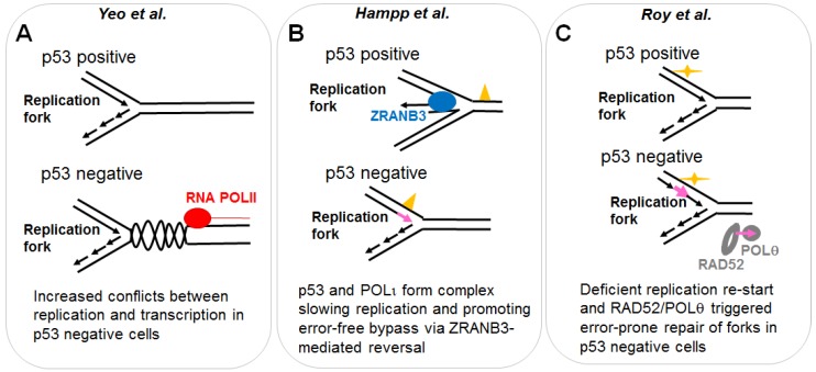
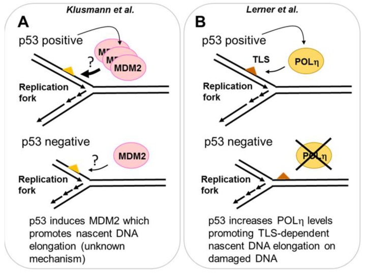
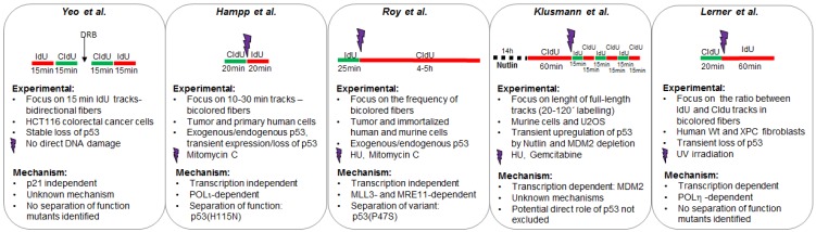
The tumor suppressor p53 is a transcriptional factor broadly mutated in cancer. Most inactivating and gain of function mutations disrupt the sequence-specific DNA binding domain, which activates target genes. This is perhaps the main reason why most research has focused on the relevance of such transcriptional activity for the prevention or elimination of cancer cells. Notwithstanding, transcriptional regulation may not be the only mechanism underlying its role in tumor suppression and therapeutic responses. In the past, a direct role of p53 in DNA repair transactions that include the regulation of homologous recombination has been suggested. More recently, the localization of p53 at replication forks has been demonstrated and the effect of p53 on nascent DNA elongation has been explored. While some data sets indicate that the regulation of ongoing replication forks by p53 may be mediated by p53 targets such as MDM2 (murine double minute 2) and polymerase (POL) eta other evidences demonstrate that p53 is capable of controlling DNA replication by directly interacting with the replisome and altering its composition. In addition to discussing such findings, this review will also analyze the impact that p53-mediated control of ongoing DNA replication has on treatment responses and tumor suppressor abilities of this important anti-oncogene.

Keywords: MRE11; POL iota; POL teta; RAD52; ZRANB3; fork reversal; mutant p53; template switching; therapy resistance; translesion DNA synthesis.


Conflict of interest statement

The authors declare no conflicts of interest.

Figures

Figure 1
Figure 1
DNA spreading assay and protocols used to study the role of p53 in the regulation of DNA replication choreography. (A) Asynchronous cells are subjected to two subsequent incorporations of thymidine analogs (CldU and IdU). Immediately after the second pulse, samples are collected and harvested in denaturing conditions on a tilted coverslip. Cells are lysed while their content scrolls down the coverslip and DNA is spread on its surface. CldU and IdU are detected using specific antibodies. DNA track that incorporated the thymidine analogs are visualized as bicolored fibers. Detailed protocols can be found as described previously [25]. By determining the length of each track, a number of DNA replication parameters can be revealed. Examples that will be discussed in this review include (B) changes in the speed of the tracks which are usually associated with the accumulation of DNA lesions in the template DNA [26]; (C) the average speed for each track; (D) the frequency of origin firing defined as the frequency of origins fired during first and the second analog incorporation [27]; (E) the efficiency of replication restart, estimated by combining the second label with a replication blocking agent such as the inhibitor of ribonucleotide reductase, hydroxyurea. Different protocols were applied in the literature [28], but in the context of p53 a modified version of the restart protocol was used as detailed in this figure [22].
Figure 2
Figure 2
A potential direct role of p53 in DNA replication. The role of p53 in DNA replication was evaluated using the DNA spreading technology. (A) p53 prevents the generation and/or the stabilization of topological conflicts between transcription and DNA replication. The mechanism by which p53 prevents such conflicts is unknown. p53 deficient cells, which accumulate such type of conflicts on replicating DNA, rely heavily on Topo2. Inhibitors of such enzyme, selectively kill p53 deficient cells. (B) p53 positive cells are more prone to the activation of an error-free post-replication repair mechanism resulting in replication associated recombination, which likely depends on ZRANB3 mediated fork reversal [39]. Such an effect correlates with a reduction in nascent DNA elongation speed in p53 expressing cells and depends on the specialized DNA POLι, which interacts with p53. An H115N mutant of p53, impaired in its exonuclease activity, is incapable of interacting with POLι, promoting replication-associated recombination and restraining nascent DNA elongation. Yellow triangle: DNA damage caused by MMC. Pink arrow: Mutagenic DNA replication events, possibly dependent on specialized POLs. (C) p53 promotes DNA replication continuity and prevents the recruitment of RAD52 and POLθ. According to the literature RAD52 and POLθ should mediate error-prone repair of collapsed forks [49]. The use of H115N and S47P variants of p53 demonstrated that the control of DNA replication restart by p53 correlates with its tumor suppressor function independently of transcriptional activation. Yellow mark: HU treatment. Pink arrow: Mutagenic replication by POLθ and RAD52.
Figure 3
Figure 3
Transcription dependent role of p53 in DNA replication. (A) Using Nutlin-3a as an inducer of p53, a novel role of increased p53 levels on DNA replication was found. When Nutlin-3a treatment was combined with a source of replication stress (HU or gemcitabine), a role of MDM2 in the elongation of nascent DNA was revealed. Strikingly, in p53 negative cells, the MDM2 pathway that controls DNA replication is still active, despite the lack of p53-dependent induction of MDM2. The elimination of MDM2 in p53 negative cells reduces DNA elongation in such genetic background. (B) p53 induces the specialized POLη after UV irradiation. Cells with increased levels of POLη (generated by a pre-irradiation with low dose of UV) promote increased nascent DNA elongation. In p53 deficient cells, the p53 dependent induction of POLη is lost, nascent DNA elongation post-UV is impaired and the cell viability is reduced.
Figure 4
Figure 4
Different protocols used to explore the effect of p53 on DNA replication. The experimental design of each study is illustrated in the upper part of each panel. Other experimental settings are detailed in the mid-section. Mechanistic insights collected in each study are detailed in the bottom part of each panel.

References

    1. Levine A.J., Oren M. The first 30 years of p53: Growing ever more complex. Nat. Rev. Cancer. 2009;9:749–758. doi: 10.1038/nrc2723. - DOI - PMC - PubMed
    1. Joerger A.C., Fersht A.R. The p53 pathway: Origins, inactivation in cancer, and emerging therapeutic approaches. Annu. Rev. Biochem. 2016;85:375–404. doi: 10.1146/annurev-biochem-060815-014710. - DOI - PubMed
    1. Cho Y., Gorina S., Jeffrey P.D., Pavletich N.P. Crystal structure of a p53 tumor suppressor-DNA complex: Understanding tumorigenic mutations. Science. 1994;265:346–355. doi: 10.1126/science.8023157. - DOI - PubMed
    1. Mello S.S., Attardi L.D. Deciphering p53 signaling in tumor suppression. Curr. Opin. Cell Biol. 2018;51:65–72. doi: 10.1016/j.ceb.2017.11.005. - DOI - PMC - PubMed
    1. Bertrand P., Saintigny Y., Lopez B.S. P53’s double life: Transactivation-independent repression of homologous recombination. Trends Genet. TIG. 2004;20:235–243. doi: 10.1016/j.tig.2004.04.003. - DOI - PubMed

LinkOut - more resources