

Progression of Geographic Atrophy in Age-related Macular Degeneration: AREDS2 Report Number 16
- PMID: 30060980
- PMCID: PMC6246813
- DOI: 10.1016/j.ophtha.2018.05.028
Progression of Geographic Atrophy in Age-related Macular Degeneration: AREDS2 Report Number 16
Abstract
Purpose: To analyze the prevalence, incidence, and clinical characteristics of eyes with geographic atrophy (GA) in age-related macular degeneration (AMD), including clinical and genetic factors affecting enlargement.
Design: Prospective cohort study within a controlled clinical trial.
Participants: Age-Related Eye Disease Study 2 (AREDS2) participants, aged 50-85 years.
Methods: Baseline and annual stereoscopic color fundus photographs were evaluated for GA presence and area. Analyses included GA prevalence and incidence rates, Kaplan-Meier rates, mixed-model regression, and multivariable analysis of the square root of GA, area adjusted for covariates, including clinical/imaging characteristics and genotype.
Main outcome measures: (1) Presence or development of GA; (2) change in the square root of GA area over time.
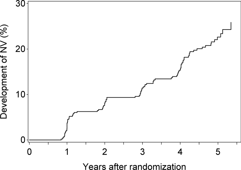
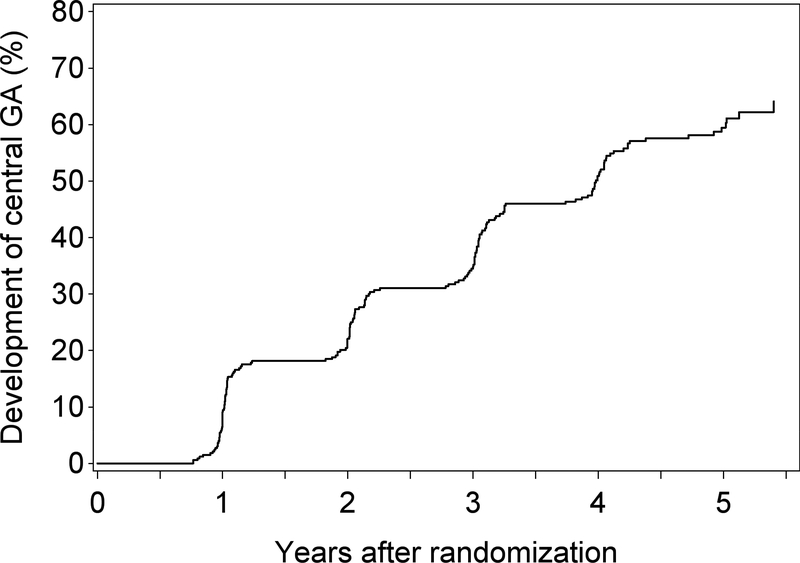
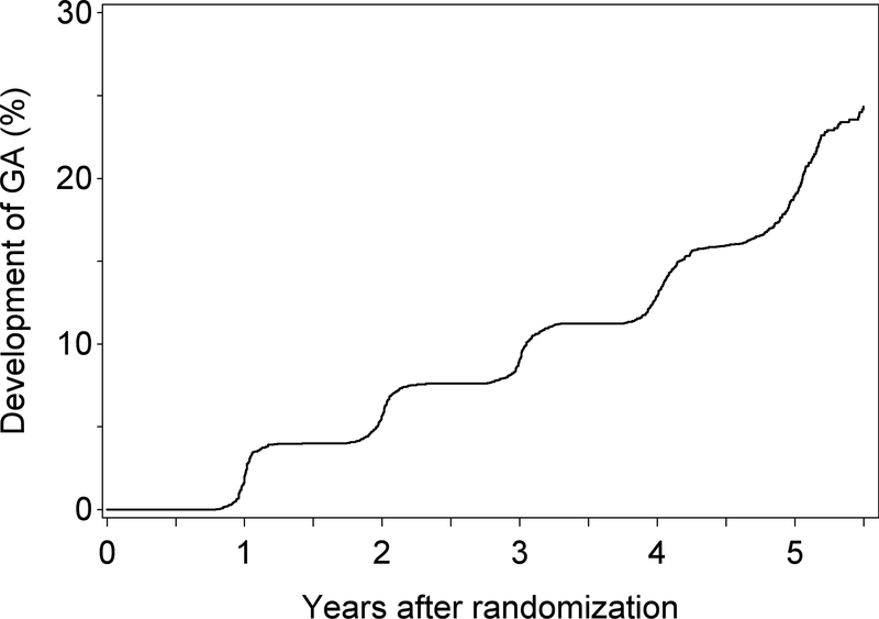
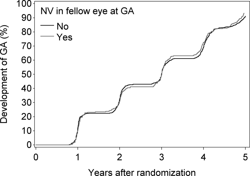
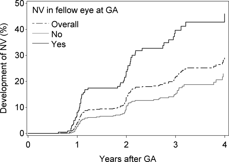
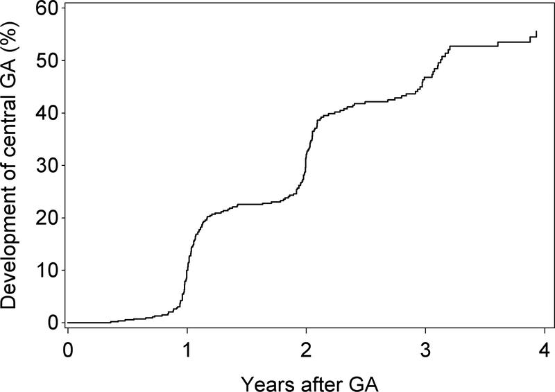
Results: At baseline, 517 eyes (6.2%) of 411 participants (9.8%) had pre-existing GA (without neovascular AMD), with the following characteristics: 33% central, 67% noncentral; and the following configurations: 36% small, 26% solid/unifocal, 24% multifocal, 9% horseshoe/ring, and 6% indeterminate. Of the remaining 6530 eyes at risk, 1099 eyes (17.3%) of 883 participants developed incident GA without prior neovascular disease during mean follow-up of 4.4 years. The Kaplan-Meier rate of incident GA was 19% of eyes at 5 years. In eyes with incident GA, 4-year risk of subsequent neovascular AMD was 29%. In eyes with incident noncentral GA, 4-year risk of central involvement was 57%. GA enlargement rate (following square root transformation) was similar in eyes with pre-existing GA (0.29 mm/year; 95% confidence interval 0.27-0.30) and incident GA (0.28 mm/year; 0.27-0.30). In the combined group, GA enlargement was significantly faster with noncentrality, multifocality, intermediate baseline size, and bilateral GA (P < 0.0001 for interaction in each case) but not with AREDS2 treatment assignment (P = 0.33) or smoking status (P = 0.05). Enlargement was significantly faster with ARMS2 risk (P < 0.0001), C3 non-risk (P = 0.0002), and APOE non-risk (P = 0.001) genotypes.
Conclusions: Analyses of AREDS2 data on natural history of GA provide representative data on GA evolution and enlargement. GA enlargement, which was influenced by lesion features, was relentless, resulting in rapid central vision loss. The genetic variants associated with faster enlargement were partially distinct from those associated with risk of incident GA. These findings are relevant to further investigations of GA pathogenesis and clinical trial planning.
Published by Elsevier Inc.
Conflict of interest statement
Conflicts of Interest:
Tiarnan Keenan, Elvira Agrón, Amitha Domalpally, Traci Clemons, PhD Freekje van Asten, Wai Wong, Emily Chew, Rinki Ratnapriya, Michael Klein, Frederick Ferris: None.
Ronald P. Danis: Consultant to Ionis Pharmaceuticals and KangHong Pharmaceuticals and is an employee and an equity owner of EyeKor, Inc
SriniVas Sadda: Consultant to Allergan, Genentech, Novartis, Thrombogenics, Iconic, NightStar, Centervue, Heidelberg, Optos and receives research support from Genentech, Carl Zeiss Meditec, Optos.
Philip Rosenfeld: Consultant to Acuela, Apellis Boehringer-Ingelheim, Carol Zeiss Meditec, Cell Cure Neurosciences, Chengdu Kanghong Biotech, Isarna Therapeutics, Genetech, Healios K.K., Hemera Biosciences, F. Hoffman-LaRoche Ltd, Ocudyne, Ocunexus, Tyrogenex, Unity Biotechnology: receives research support: Astellas Institute for Regenerative Medicine (AIRM), Carl Zeiss Medictec, Genentech, Tyrogenex; and has equity interest in Apellis, Digisight, Ocudyne.
Figures
Comment in
-
Re: Keenan et al.: Progression of geographic atrophy in age-related macular degeneration (Ophthalmology. 2018;125:1913-1928).Ophthalmology. 2019 May;126(5):e39-e40. doi: 10.1016/j.ophtha.2018.11.029. Ophthalmology. 2019. PMID: 31005199 No abstract available.
-
Reply.Ophthalmology. 2019 May;126(5):e40-e41. doi: 10.1016/j.ophtha.2018.11.030. Ophthalmology. 2019. PMID: 31005200 No abstract available.
References
-
- Gass JD. Drusen and disciform macular detachment and degeneration. Arch Ophthalmol. 1973;90(3):206–17. - PubMed
-
- AREDS RG. The Age-Related Eye Disease Study system for classifying age-related macular degeneration from stereoscopic color fundus photographs: the Age-Related Eye Disease Study Report Number 6. Am J Ophthalmol. 2001;132(5):668–81. - PubMed
Publication types
MeSH terms
Substances
Grants and funding
LinkOut - more resources
Full Text Sources
Other Literature Sources
Medical
Molecular Biology Databases
Miscellaneous
