. 2018 Sep 5;26(9):2189-2205.
doi: 10.1016/j.ymthe.2018.07.009. Epub 2018 Jul 12.

MicroRNA-218-5p Promotes Endovascular Trophoblast Differentiation and Spiral Artery Remodeling

Affiliations

MicroRNA-218-5p Promotes Endovascular Trophoblast Differentiation and Spiral Artery Remodeling

Jelena Brkić et al. Mol Ther. .

Abstract

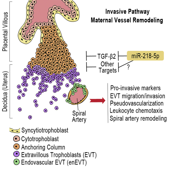
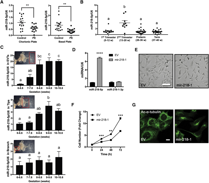
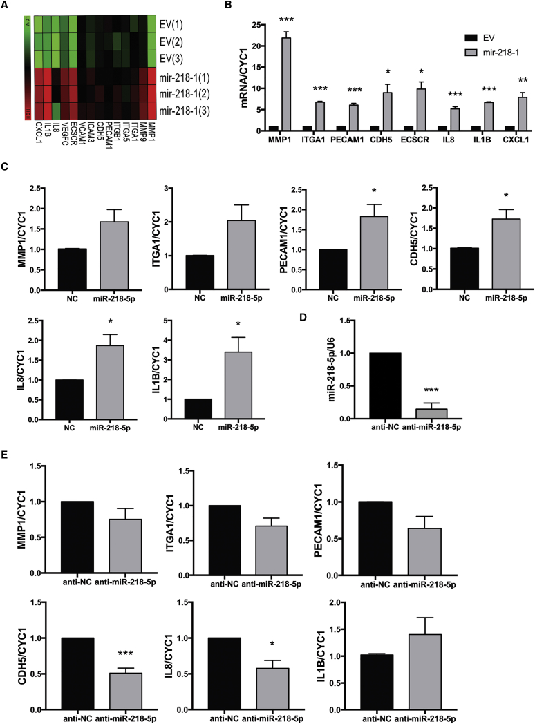
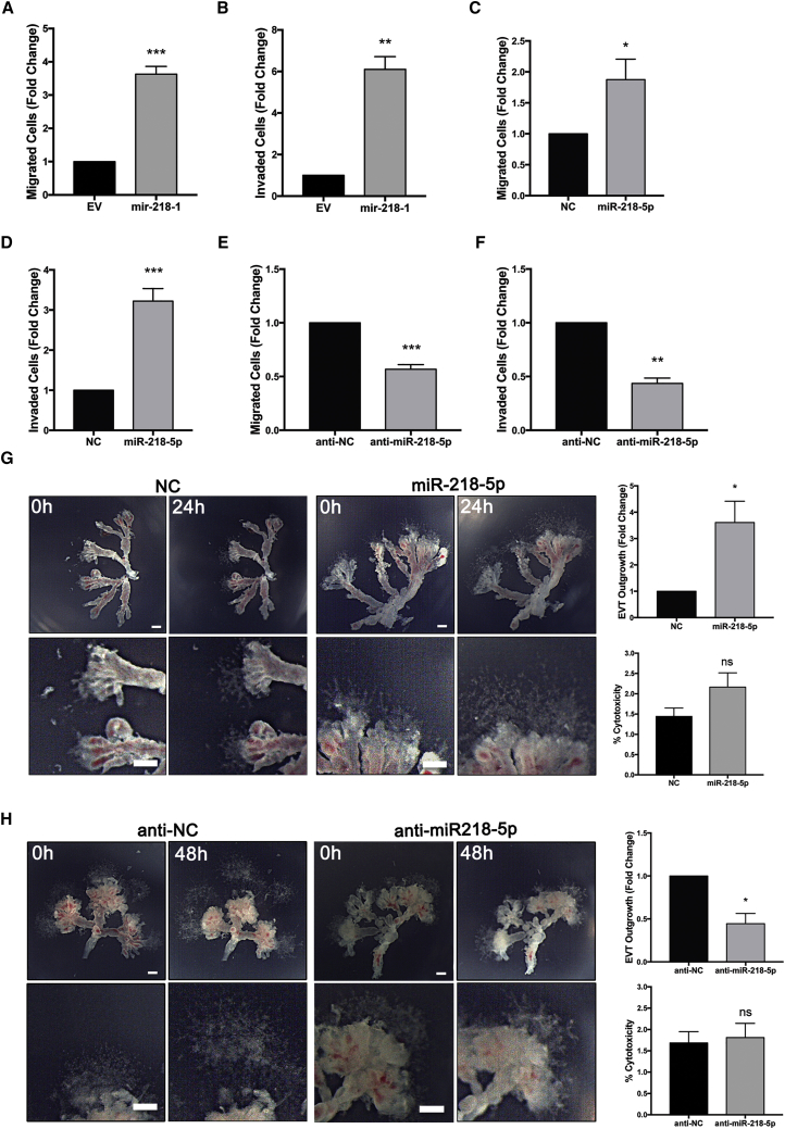
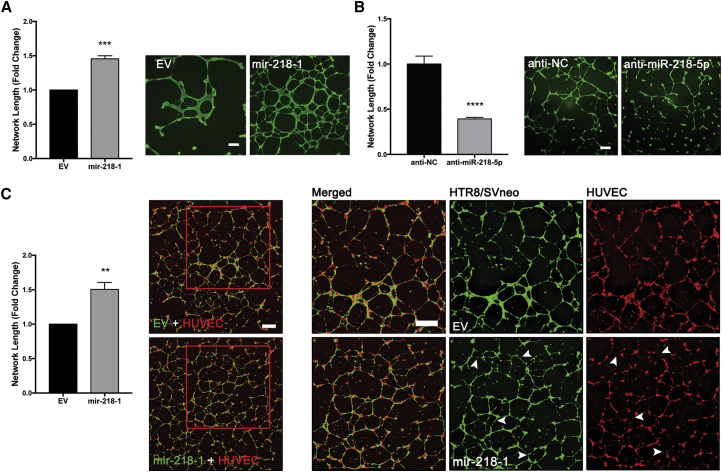
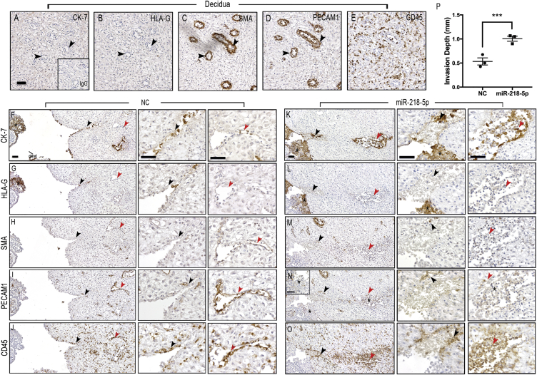
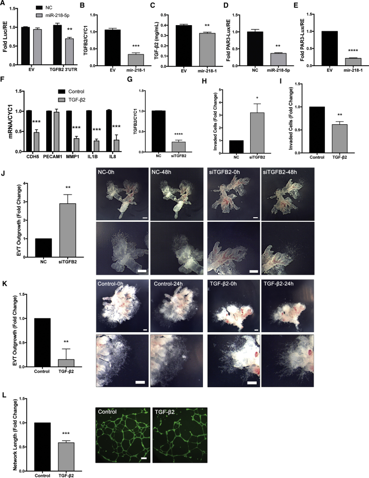
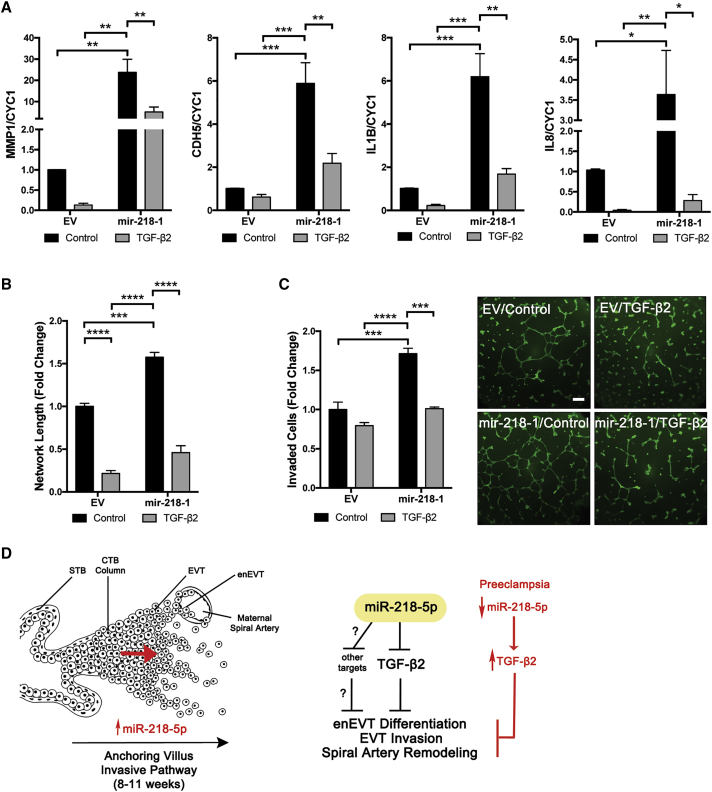
Preeclampsia (PE) is the leading cause of maternal and neonatal morbidity and mortality. Defects in trophoblast invasion, differentiation of endovascular extravillous trophoblasts (enEVTs), and spiral artery remodeling are key factors in PE development. There are no markers clinically available to predict PE, leaving expedited delivery as the only effective therapy. Dysregulation of miRNA in clinical tissues and maternal circulation have opened a new avenue for biomarker discovery. In this study, we investigated the role of miR-218-5p in PE development. miR-218-5p was highly expressed in EVTs and significantly downregulated in PE placentas. Using first-trimester trophoblast cell lines and human placental explants, we found that miR-218-5p overexpression promoted, whereas anti-miR-218-5p suppressed, trophoblast invasion, EVT outgrowth, and enEVT differentiation. Furthermore, miR-218-5p accelerated spiral artery remodeling in a decidua-placenta co-culture. The effect of miR-218-5p was mediated by the suppression of transforming growth factor (TGF)-β2 signaling. Silencing of TGFB2 mimicked, whereas treatment with TGF-β2 partially reversed, the effects of miR-218-5p. Taken together, these findings demonstrate that miR-218-5p promotes trophoblast invasion and enEVT differentiation through a novel miR-218-5p-TGF-β2 pathway. This study elucidates the role of an miRNA in enEVT differentiation and spiral artery remodeling and suggests that downregulation of miR-218-5p contributes to PE development.

Keywords: TGF-β; endovascular trophoblasts; miR-218-5p; micorRNA; placenta-decidua co-culture; preeclampsia; remodeling; spiral artery; vascular transformation.


Figures

None
Graphical abstract
Figure 1
Figure 1
Expression Pattern of miR-218-5p in Human Healthy and PE Placentas (A) miR-218-5p expression in normal and PE placentas. PE and healthy term placentas between 35 and 39 weeks of gestation were collected from the basal and chorionic plates (n = 15 placentas). miR-218-5p was downregulated in PE placentas compared with the control. (B) miR-218-5p levels in placentas across gestation (n = 9–24 placentas). An increase (p < 0.0001) in miR-218-5p levels during the second trimester was observed compared with all other gestational periods. Different letters above bars denote statistical significance. (C) miR-218-5p expression in different regions of anchoring villi during early pregnancy. Placental samples were obtained from elective terminations from 6–10.6 weeks gestation, dissected into EVTs (top), tips (center), and branches (bottom), and measured for miR-218-5p levels. Different letters above bars denote statistical significance (n = 3–4 placentas). An increase in miR-218-5p was observed from 8 and 9 weeks in EVTs and tips, respectively. No difference in miR-218-5p expression was observed in villi branches. (D) Confirmation of miR-218-5p overexpression in HTR-8/SVneo cells stably transfected with mir-218-1 versus cells transfected with empty vector (EV). (E) Morphological examination of stable mir-218-1 and control cells seeded in equal numbers showed that mir-218-1 cells were smaller and spindle-shaped, with less cell-cell contact compared with EV cells. Scale bar, 100 μm. (F) Cell proliferation assays showed mir-218-1-overexpressing cells grew slower than EV cells. (G) Immunofluorescence staining of acetylated α-tubulin. mir-218-1 cells displayed a more disorganized microtubule network and asymmetric organization. Scale bar, 10 μm. All cell data are representative of three independent experiments. Statistical analysis was performed using (A) unpaired two-tailed Student’s t test with Welch’s correction, (B and C) one-way ANOVA with Tukey’s test, or (D and F) multiple t tests. **p < 0.01, ***p < 0.001, ****p < 0.0001. Error bars represent SEM.
Figure 2
Figure 2
miR-218-5p Increases the Expression of Key Markers of Trophoblast Invasion and Endovascular Trophoblast Differentiation (A) Gene expression profiling in EV and mir-218-1 stable cells by cDNA microarray. Several markers of trophoblast differentiation and genes involved in invasion were greatly upregulated in mir-218-1 cells. (B) Validation of genes regulated by mir-218-1. All markers were significantly upregulated in mir-218-1 cells (n = 3 experiments). (C) Regulation of gene expression by miR-218-5p in first-trimester placental explants. Placental tissues were dissected to enrich extravillous trophoblast (EVT) columns and treated with miR-218-5p or a scramble non-targeting control (NC) for 48 hr. miR-218-5p significantly upregulated PECAM1, CDH5, IL-8, and IL-1B mRNA levels versus NC (n = 6 placentas). (D) Anti-miR-218-5p inhibits endogenous miR-218-5p in placental explants. Placental tissues were incubated with anti-miR-218-5p for 48 hr, and miR-218-5p levels were measured (n = 6 placentas). miR-218-5p was downregulated by anti-miR-218-5p. (E) Anti-miR-218-5p downregulated marker gene expression. The RNA samples described in (D) were used for real-time qPCR. Although most markers measured showed a trend of decrease, only CDH5 and IL-8 were significant. Statistical analysis was performed by (B) multiple t tests (α = 0.05) corrected for multiple comparisons with the Holm-Sidak method and (C and D) Welch’s t test (p < 0.05, 95% confidence interval [CI]) using GraphPad Prism. *p < 0.05, **p < 0.01, ***p < 0.001. Error bars represent SEM.
Figure 3
Figure 3
miR-218-5p Promotes Trophoblast Invasion, Migration, and EVT Outgrowth (A–F) Cells were seeded on transwells without and with Matrigel to assess cell migration or invasion, respectively. Stable transfection of mir-218-1 increased cell migration (A) and invasion (B) compared with control cells transfected with EV. Transient transfection of the miR-218-5p mimics also promoted cell migration (C) and invasion (D) compared with NC. Conversely, anti-miR-218-5p significantly decreased the migratory (E) and invasive (F) ability of the cells compared with controls. The results presented in (A)–(F) are data pooled from a minimum of three independent experiments. (G) Explants from first-trimester placentas were placed on Matrigel and treated with miR-218-5p or control (200 nM) for 24 hr. EVT outgrowth was measured at the time of treatment (0 hr) and at termination of the experiment (24 hr). An increase in EVT outgrowth was observed in miR-218-5p-treated tissues. (H) Explant tissues were treated with anti-miR-218-5p or the control (200 nM) for 48 hr. A decrease in EVT outgrowth was observed with anti-miR-218-5p treatment. Neither miR-218-5p (G) nor anti-miR-218-5p (H) had significant effects on cytotoxicity. Representative images are shown, and bar graphs present data from four experiments with unique placentas. *p < 0.05, **p < 0.01, ***p < 0.001. Error bars represent SEM. Scale bars, 250 μm.
Figure 4
Figure 4
Overexpression of mir-218-1 Promotes Endothelium-like Network Formation (A) mir-218-1 enhances network formation. Control and mir-218-1 stable cells were seeded on Matrigel-coated wells, and network formation was assessed 18 hr after seeding. mir-218-1 cells displayed an increased ability to align into network structures compared with control cells (n = 3 experiments). A representative image is shown. (B) Anti-miR-218-5p inhibits network formation. HTR8/SVneo cells were transiently transfected with anti-miR-218-5p, and network formation assays were performed. Compared with an NC oligo (anti-NC), cells transfected with anti-miR-218-5p showed a decreased ability to form network structures (n = 3). One representative experiment is shown. (C) mir-218-1 stimulates network formation in a co-culture of trophoblasts and HUVECs. Control (EV) or mir-218-1 stable trophoblasts (green) were seeded on Matrigel at a one-to-one ratio with HUVECs (red), and cells were allowed to co-localize and form networks for 18 hr. mir-218-1-HUVEC co-culture showed a more complex network with a larger total length (n = 3). Examination of networks formed on the left showed that, in co-culture with control (EV) trophoblasts, HUVECs formed intact branches. However, in co-culture with mir-218-1-overexpressing cells, the networks formed by HUVECs were not intact (white arrowheads). Representative images are shown. **p < 0.01, ***p < 0.001, ****p < 0.0001. Error bars represent SEM. Scale bars, 500 μm.
Figure 5
Figure 5
miR-218-5p Accelerates Spiral Artery Remodeling (A–E) Investigation of markers for trophoblasts (CK-7 and HLA-G), spiral arteries (SMA and PECAM1), and leukocytes (CD45) in decidua parietalis cultured alone. The tissue was negative for CK-7 and HLA-G (A and B, black arrowheads). Tight, non-invaded arterioles stained positive for SMA and PECAM1 (C and D, black arrowheads). Lymphocytes positive for CD45 were spread evenly throughout the tissue (E). (F–J) Placenta tissue pre-treated with NC was assessed for the degree of vessel remodeling. EVTs positive for CK-7 (F) and HLA-G (G) (black asterisks, *) entered the proximal arteriole (black arrowheads), and a few cells also reached the distal portion (red arrowheads). Smooth muscle (H) and endothelial (I) cells were mostly removed in the proximal portion (black arrowheads) of the spiral arteriole but remained intact in the distal portion (red arrowheads), suggesting that the vessel was at an early “active phase” of vascular transformation. Leukocyte recruitment (J) at the site of remodeling was observed (arrowheads). (K–O) Placenta tissue pre-treated with miR-218-5p showed an accelerated degree of vessel remodeling. CK7-positive (K) and HLA-G-positive (L) EVTs (black asterisks, *) entered the proximal portion of the arteriole (black arrowheads) and invaded the distal portion (red arrowheads). Only a few cells positive for SMA (M) remained at the proximal (black arrowheads) but also the distal (red arrowheads) portions of the arteriole. Smooth muscle cells in both proximal (black arrowheads) and distal portions had been predominantly removed, and cells in the lumen showed signs of phagocytosis (red arrowheads). Only residual PECAM1 -positive endothelial cells (N) remained at both the proximal (black arrowheads) and distal (red arrowheads) portions of arteriole. EVTs were weakly positive for PECAM1 in the placenta column and the endovascular EVTs at the distal site of remodeling (black asterisks, *). Extensive recruitment of leukocytes to the site of active remodeling (arrowheads) and associated clearing of leukocytes from surrounding tissue was observed (O). (P) Quantification of the remodeling arterioles showed a doubling in depth of trophoblast invasion and vessel remodeling in miR-218-5p-pretreated placentas compared with the control (n = 3 independent experiments with 2–3 co-cultures per experiment). Error bars represent SEM. Scale bars, 50 μm.
Figure 6
Figure 6
miR-218-5p Targets TGFB2 (A) Luciferase reporter assay. miR-218-5p reduced luciferase activity in cells transfected with the TGFB2 3′ UTR reporter but not in cells transfected with the EV (n = 3). (B) TGFB2 mRNA was downregulated in mir-218-1 cells compared with the control (n = 3). (C) ELISA quantification of TGF-β2 showed a downregulation in the conditioned media collected from mir-218-1 stable cells compared with EV (n = 3). (D) Transient transfection of miR-218-5p reduced SMAD transcription activity, as measured using a SMAD response reporter, pAR3-lux (n = 3). (E) mir-218-1-overexpressing cells had lower SMAD-induced luciferase activity compared with control cells (n = 3). (F) Cells treated with recombinant human TGF-β2 (10 ng/mL) for 24 hr downregulated key markers of invasion and endovascular EVT differentiation (n = 3). (G) Validation of siRNA targeting TGFB2 (siTGFB2) (n = 3). (H) Silencing of TGFB2 increased cell invasion (n = 3). (I) Treatment with TGF-β2 suppressed invasion (n = 3). (J) Silencing of TGFB2 enhanced first-trimester placental explant outgrowth. Placental explants were treated with 200 nM siTGFB2 or NC for 48 hr (n = 4). (K) Treatment with TGF-β2 (10 ng/mL) for 24 hr inhibited placental explant outgrowth (n = 5). (L) TGF-β2 inhibits network formation. HTR8/SVneo cells pre-treated with TGF-β2 (10 ng/mL) showed decreased network structures on Matrigel compared with the control (n = 3). Statistical analysis was performed using a two-tailed unpaired t test or (F) multiple t tests corrected for multiple comparisons with the Holm-Sidak method. *p < 0.05, **p < 0.01, ***p < 0.001, ****p < 0.0001. Error bars represent SEM. Scale bars, 250 μm.
Figure 7
Figure 7
miR-218-5p Exerts Its Effect through Suppression of TGF-β2 (A) TGF-β2 reversed the effect of mir-218-1 on gene expression. EV control and mir-218-1-overexpressing stable cells were treated with TGF-β2 (10 ng/mL). mir-218-1 increased MMP-1, CDH5, IL-1B, and IL-8 mRNA levels, but these effects were abolished by TGF-β2. (B) TGF-β2 reversed the effect of mir-218-1 on cell invasion. Treatment with TGF-β2 inhibited the promoting effect of mir-218-1 on cell invasion. (C) TGF-β2 reversed the effect of mir-218-1 on network formation. TGF-β2 treatment reduced the ability of mir-218-1 stable cells to form network-like structures. (D) Proposed role of miR-218-5p in placental development and in the pathogenesis of preeclampsia. miR-218-5p is upregulated in EVTs to promote trophoblast invasion, endovascular differentiation, and spiral artery remodeling, in part through suppression of TGF-β2 signaling. Other target genes remain to be identified. Downregulation of miR-218-5p may lead to upregulation of TGF-β2 and other genes, which contributes to the shallow invasion of EVTs and defective spiral artery remodeling observed in PE placentas. STB, syncytiotrophoblast; CTB, cytotrophoblast; EVT, extravillous trophoblast; enEVT, endovascular EVT. *p < 0.05, **p < 0.01, ***p < 0.001, ****p < 0.0001. Error bars represent SEM. Scale bar, 500 μm.

References

    1. Duley L. The global impact of pre-eclampsia and eclampsia. Semin. Perinatol. 2009;33:130–137. - PubMed
    1. Backes C.H., Markham K., Moorehead P., Cordero L., Nankervis C.A., Giannone P.J. Maternal preeclampsia and neonatal outcomes. J. Pregnancy. 2011;2011:214365–214367. - PMC - PubMed
    1. Neiger R. Long-Term Effects of Pregnancy Complications on Maternal Health: A Review. J. Clin. Med. 2017;6:76. - PMC - PubMed
    1. Riise H.K.R., Sulo G., Tell G.S., Igland J., Nygård O., Vollset S.E., Iversen A.C., Austgulen R., Daltveit A.K. Incident Coronary Heart Disease After Preeclampsia: Role of Reduced Fetal Growth, Preterm Delivery, and Parity. J. Am. Heart Assoc. 2017;6:e004158. - PMC - PubMed
    1. Davis E.F., Lazdam M., Lewandowski A.J., Worton S.A., Kelly B., Kenworthy Y., Adwani S., Wilkinson A.R., McCormick K., Sargent I. Cardiovascular risk factors in children and young adults born to preeclamptic pregnancies: a systematic review. Pediatrics. 2012;129:e1552–e1561. - PubMed

Publication types

LinkOut - more resources