. 2018 Aug 14;115(33):8340-8345.
doi: 10.1073/pnas.1807881115. Epub 2018 Jul 30.

Identification of a biologically active fragment of ALK and LTK-Ligand 2 (augmentor-α)

Affiliations

Identification of a biologically active fragment of ALK and LTK-Ligand 2 (augmentor-α)

Andrey V Reshetnyak et al. Proc Natl Acad Sci U S A. .

Abstract

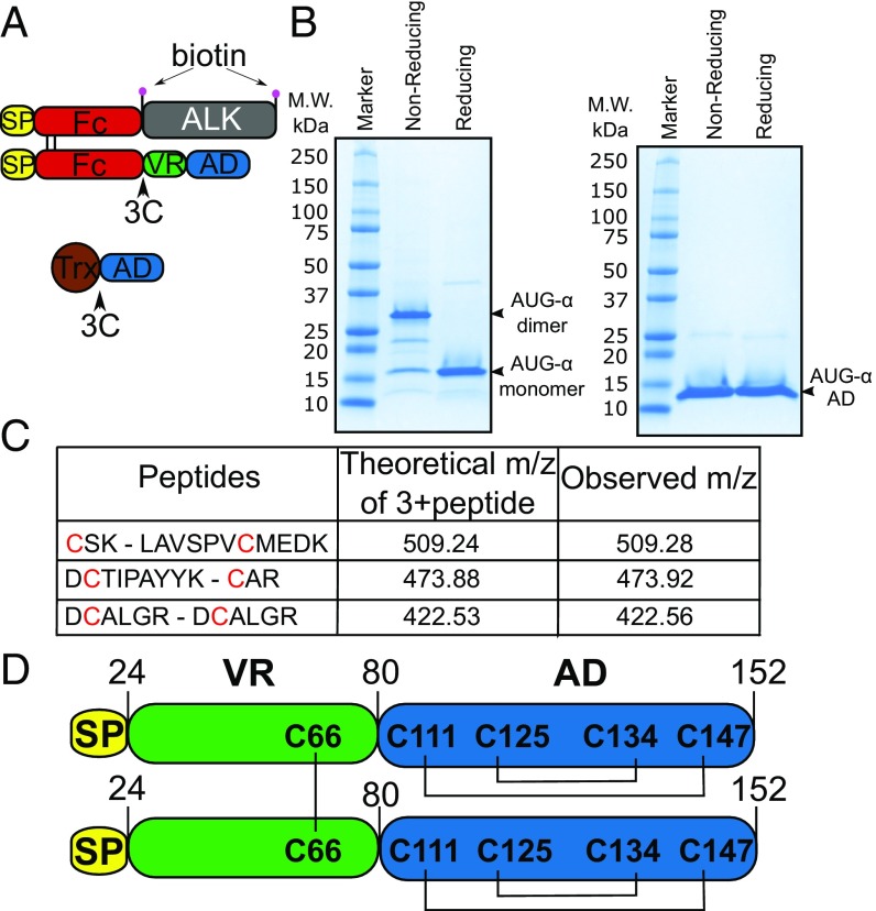
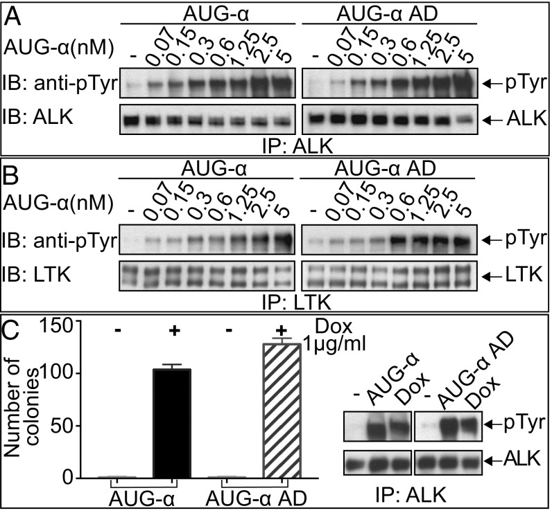
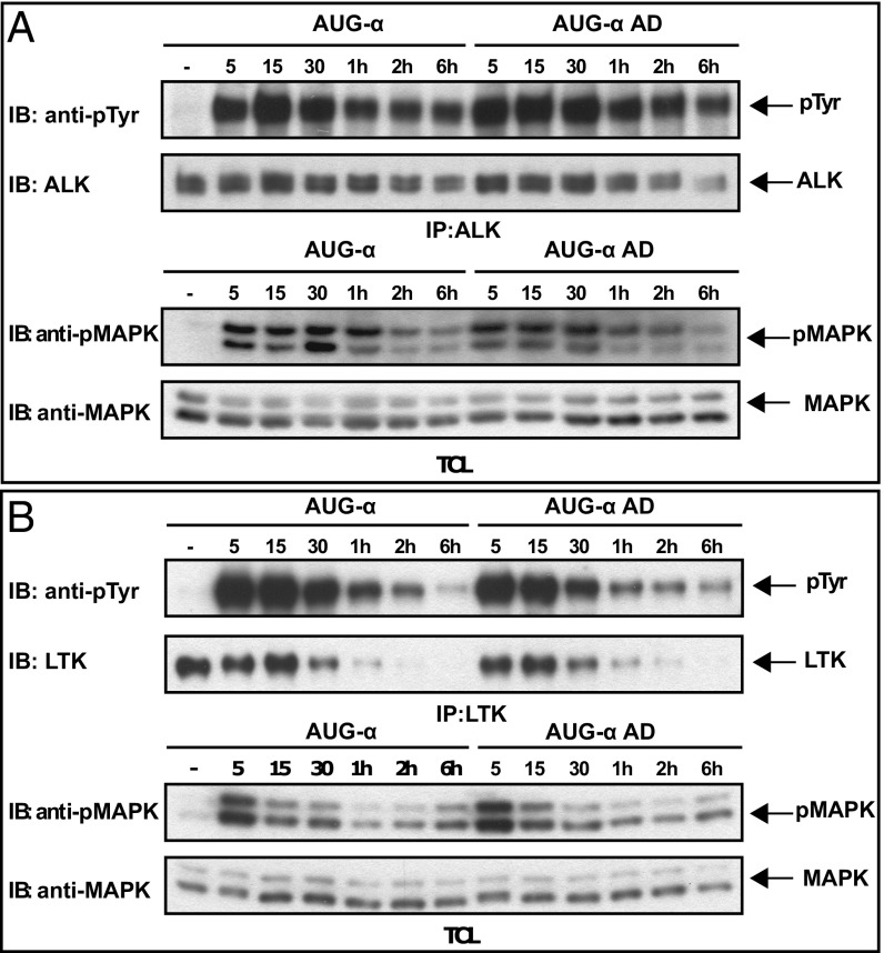
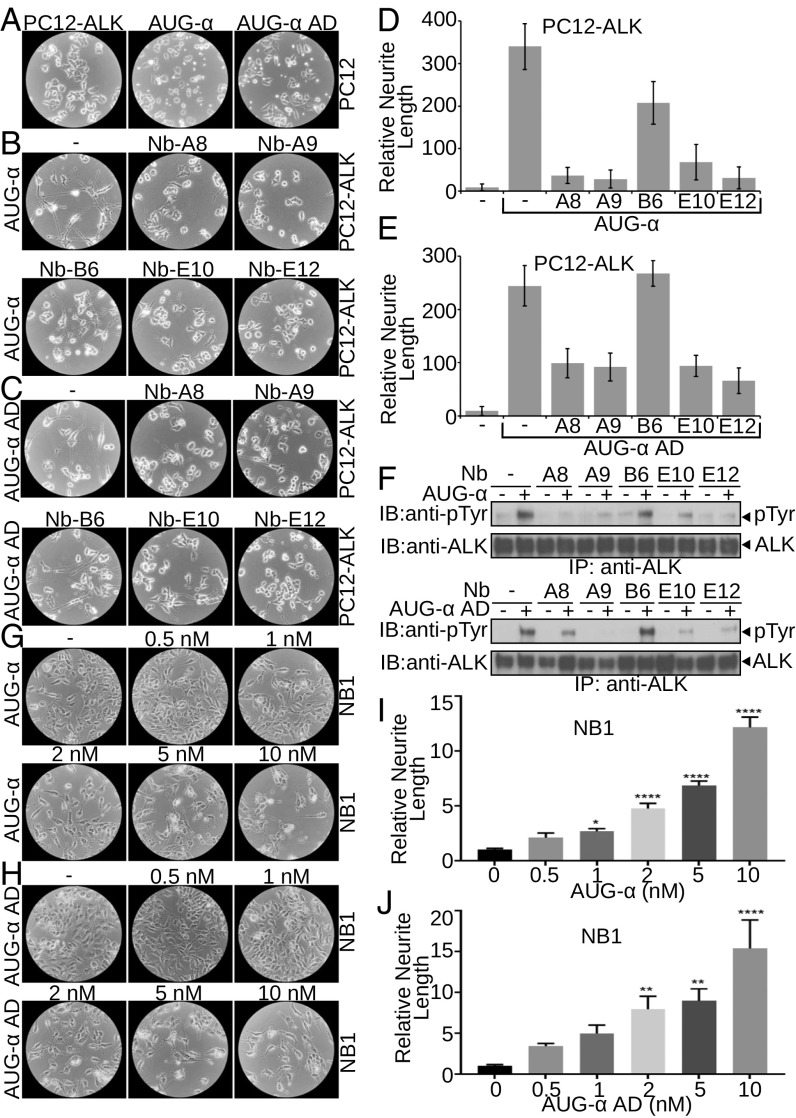
Elucidating the physiological roles and modes of action of the recently discovered ligands (designated ALKAL1,2 or AUG-α,β) of the receptor tyrosine kinases Anaplastic Lymphoma Kinase (ALK) and Leukocyte Tyrosine Kinase (LTK) has been limited by difficulties in producing sufficient amounts of the two ligands and their poor stability. Here we describe procedures for expression and purification of AUG-α and a deletion mutant lacking the N-terminal variable region. Detailed biochemical characterization of AUG-α by mass spectrometry shows that the four conserved cysteines located in the augmentor domain (AD) form two intramolecular disulfide bridges while a fifth, primate-specific cysteine located in the N-terminal variable region mediates dimerization through formation of a disulfide bridge between two AUG-α molecules. In contrast to AUG-α, the capacity of AUG-α AD to undergo dimerization is strongly compromised. However, full-length AUG-α and the AUG-α AD deletion mutant stimulate similar tyrosine phosphorylation of cells expressing either ALK or LTK. Both AUG-α and AUG-α AD also stimulate a similar profile of MAP kinase response in L6 cells and colony formation in soft agar by autocrine stimulation of NIH 3T3 cells expressing ALK. Moreover, both AUG-α and AUG-α AD stimulate neuronal differentiation of human neuroblastoma NB1 and PC12 cells in a similar dose-dependent manner. Taken together, these experiments show that deletion of the N-terminal variable region minimally affects the activity of AUG-α toward LTK or ALK stimulation in cultured cells. Reduced dimerization might be compensated by high local concentration of AUG-α AD bound to ALK at the cell membrane and by potential ligand-induced receptor-receptor interactions.

Keywords: active fragment; cell signaling; cytokine; receptor activation; receptor tyrosine kinases.


Conflict of interest statement

The authors declare no conflict of interest.

Figures

Fig. 1.
Fig. 1.
AUG-α is expressed as a disulfide bridged dimer. (A) Schematic representation of expression constructs to produce full-length AUG-α from mammalian cells (Upper) or AUG-α AD from bacteria (Lower). Signal peptide is colored in yellow, Fc fragment in red, ALK fragment (residues 648–1030) in gray, AUG-α variable region (VR) in green, augmentor domain (AD) in blue, and Trx in brown. The 3C protease cleavage site is marked by an arrow, and the biotin acceptor peptides are marked by pink circles. (B) SDS/PAGE analysis of purified full-length AUG-α (Left) and a truncated version, AUG-α AD (Right), separated under nonreducing and reducing conditions as indicated in the upper panels. Molecular weight (M.W.) marker with corresponding molecular masses is shown on the left side of the gels. Under nonreducing conditions, full-length AUG-α migrates as a dimer, but migrates as a monomer upon reduction (as indicated on the right side). (C) Mass spectrometry mapping of disulfide bridges in AUG-α. Purified AUG-α was subjected to nonreducing SDS/PAGE, and the band corresponding to the dimeric form of AUG-α was excised, digested with trypsin, and used for mass spectrometry analysis. Disulfide-linked peptides are presented in the first column, and corresponding cysteines are highlighted in red. Theoretically predicted molecular masses for the corresponding 3+peptides are listed in the second column, and the observed m/z is listed in the last column. (D) Schematic representation of full-length AUG-α. Color code is the same as in A and B. Disulfide bridges are shown by black lines.
Fig. 2.
Fig. 2.
Deletion of the N-terminal variable region of AUG-α does not affect kinase activity of ALK or LTK. (A and B) Immunoblot analysis of ALK (A) or LTK (B) autophosphorylation stimulated by different concentrations (as indicated) of purified AUG-α or AUG-α AD. NIH 3T3 cells stably expressing ALK or LTK were stimulated with increasing concentration of AUG-α or AUG-α AD for 10 min at 37 °C. Lysates of unstimulated or AUG-α–stimulated cells were subjected to immunoprecipitation (IP) using anti-ALK or anti-LTK antibodies followed by SDS/PAGE and immunoblotting (IB) with anti-pTyr (IB: pTyr) or anti-ALK or anti-LTK antibodies (as indicated). (C) NIH 3T3 cells were stably transfected with ALK and a Dox-inducible construct of AUG-α or AUG-α AD. (Right) Immunoblot analysis of ALK autophosphorylation stimulated by DMSO (indicated as by “−”) and 50 μg of purified AUG-α or AUG-α AD, or Dox. (Left) Soft agar colony formation assay. Double-stable NIH 3T3 cells were treated with either DMSO or 1 µg/mL of Dox and grown in soft agar for 2 wk. Colonies were stained with crystal violet, counted, and plotted. Each experiment was performed in triplicate, and SD was calculated and plotted for each experiment.
Fig. 3.
Fig. 3.
Time courses of ALK and LTK receptor phosphorylation stimulated by AUG-α or AUG-α AD. Time courses of ALK (A) or LTK (B) phosphorylation in L6 cells stably expressing these receptors. Phosphorylation was induced by saturating levels (10 nM) of AUG-α or AUG-α AD. Cell lysates were subjected to immunoprecipitation (IP) by anti-ALK or anti-LTK antibodies, and phosphorylation was monitored by anti-pTyr antibodies (IB: anti-pTyr). Anti-ALK or anti-LTK antibodies were used as loading controls. Total cell lysates (TCL) and anti-phosphoMAPK or anti-MAPK antibodies were used to analyze MAPK phosphorylation in response to AUG-α or AUG-α AD stimulation at different time points.
Fig. 4.
Fig. 4.
AUG-α and AUG-α AD promote neurite outgrowth of neuronal PC12 and NB1 cells through ALK phosphorylation. (AF) Neurite outgrowth of PC12 cells. (A) Unstimulated PC12 cells overexpressing ALK (Left). As a control, parental PC12 cells (two Right panels) were stimulated with 10 nM of AUG-α and AUG-α AD (as indicated). (B and C) PC12 cells overexpressing ALK were pretreated with 50 nM of anti-ALK nanobodies (A8, A9, B6, E10, and E12 as indicated) and were stimulated with 10 nM of either AUG-α (B) or AUG-α AD (C). (D and E) Relative neurite length per cell; values are means ± SEM; n = 5. (F) Inhibition of AUG-α (Upper) and AUG-α AD (Lower) stimulated ALK phosphorylation by nanobodies in PC12 cells assessed by immunoblotting (IB). (GJ) Neurite outgrowth of NB1 cells stimulated by AUG-α (G and I) or AUG-α AD (H and J). NB1 cells were assayed for neurite length with 0.0, 0.5, 1.0, 2.0, 5.0, and 10.0 nM concentration of AUG-α (G and I) or Aug-α AD (H and J). (I and J) Quantification of relative neurite length. Values are means ± SEM; n = 6. *P < 0.05, **P < 0.01, ****P < 0.0001, unpaired t test.

References

    1. Lemke G. Adopting ALK and LTK. Proc Natl Acad Sci USA. 2015;112:15783–15784. - PMC - PubMed
    1. Murray PB, et al. Heparin is an activating ligand of the orphan receptor tyrosine kinase ALK. Sci Signal. 2015;8:ra6. - PubMed
    1. Zhang H, et al. Deorphanization of the human leukocyte tyrosine kinase (LTK) receptor by a signaling screen of the extracellular proteome. Proc Natl Acad Sci USA. 2014;111:15741–15745. - PMC - PubMed
    1. Ben-Neriah Y, Bauskin AR. Leukocytes express a novel gene encoding a putative transmembrane protein-kinase devoid of an extracellular domain. Nature. 1988;333:672–676. - PubMed
    1. Morris SW, et al. Fusion of a kinase gene, ALK, to a nucleolar protein gene, NPM, in non-Hodgkin’s lymphoma. Science. 1994;263:1281–1284. - PubMed

Publication types