. 2018 Aug 14;115(33):E7871-E7880.
doi: 10.1073/pnas.1800680115. Epub 2018 Jul 30.

Histone methyltransferase Smyd1 regulates mitochondrial energetics in the heart

Affiliations

Histone methyltransferase Smyd1 regulates mitochondrial energetics in the heart

Junco S Warren et al. Proc Natl Acad Sci U S A. .

Abstract

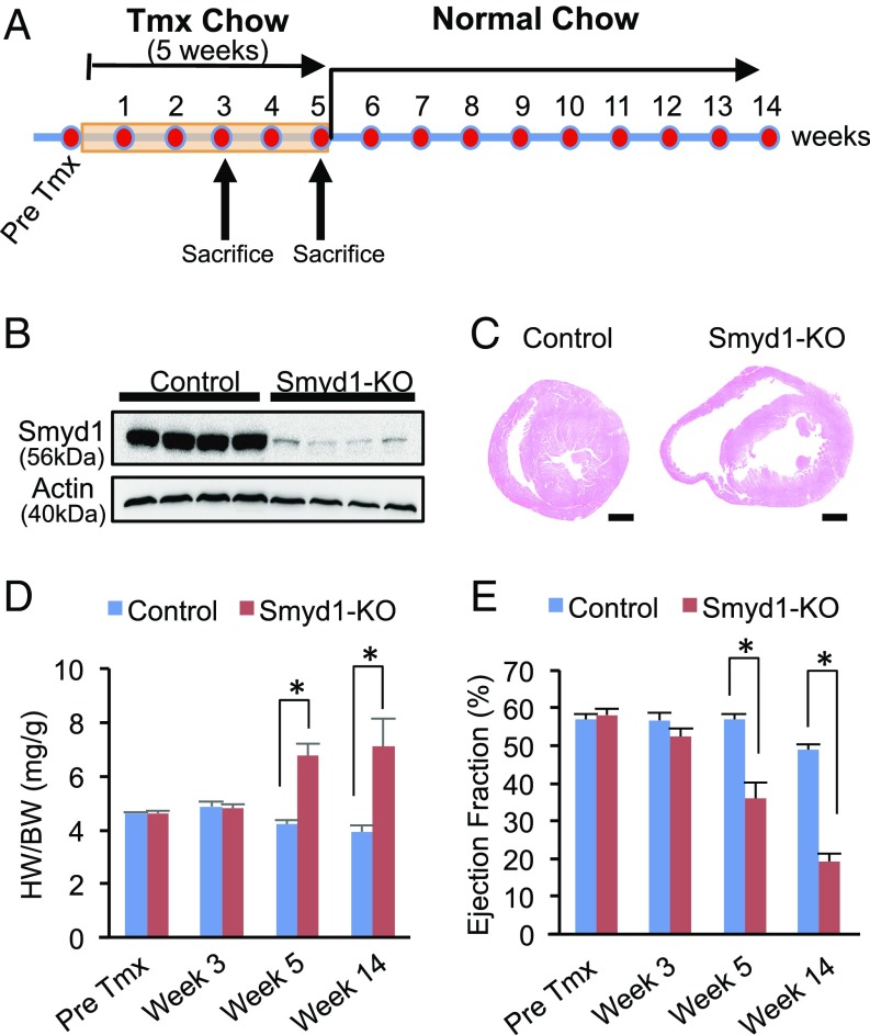
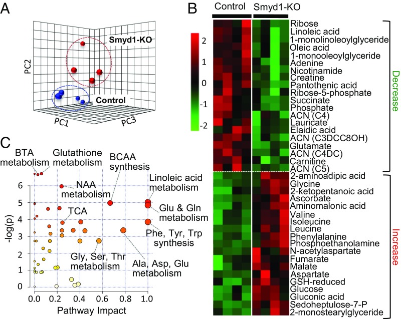
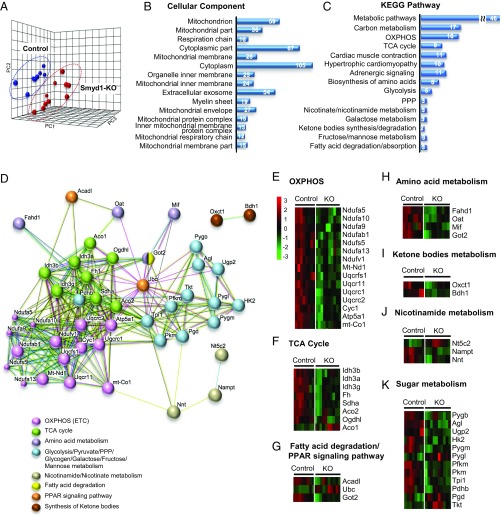
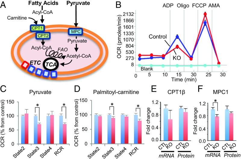
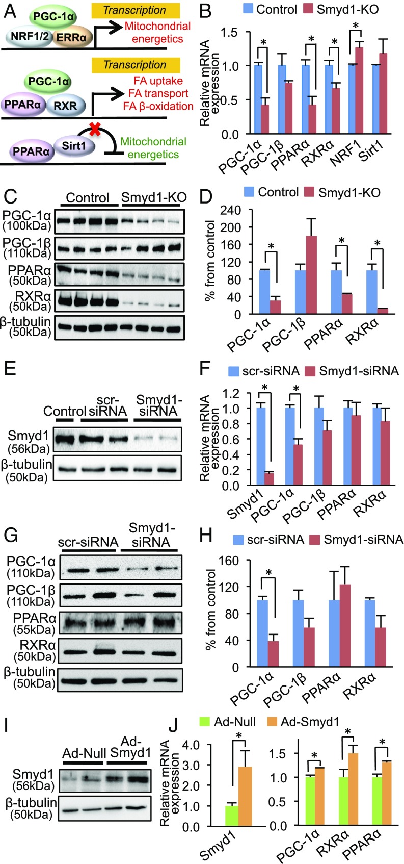
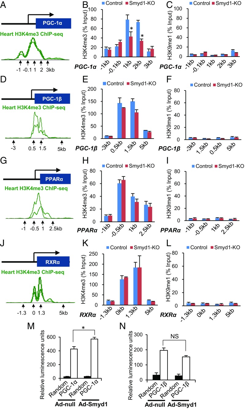
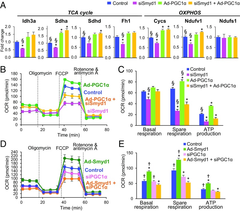
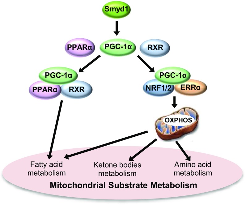
Smyd1, a muscle-specific histone methyltransferase, has established roles in skeletal and cardiac muscle development, but its role in the adult heart remains poorly understood. Our prior work demonstrated that cardiac-specific deletion of Smyd1 in adult mice (Smyd1-KO) leads to hypertrophy and heart failure. Here we show that down-regulation of mitochondrial energetics is an early event in these Smyd1-KO mice preceding the onset of structural abnormalities. This early impairment of mitochondrial energetics in Smyd1-KO mice is associated with a significant reduction in gene and protein expression of PGC-1α, PPARα, and RXRα, the master regulators of cardiac energetics. The effect of Smyd1 on PGC-1α was recapitulated in primary cultured rat ventricular myocytes, in which acute siRNA-mediated silencing of Smyd1 resulted in a greater than twofold decrease in PGC-1α expression without affecting that of PPARα or RXRα. In addition, enrichment of histone H3 lysine 4 trimethylation (a mark of gene activation) at the PGC-1α locus was markedly reduced in Smyd1-KO mice, and Smyd1-induced transcriptional activation of PGC-1α was confirmed by luciferase reporter assays. Functional confirmation of Smyd1's involvement showed an increase in mitochondrial respiration capacity induced by overexpression of Smyd1, which was abolished by siRNA-mediated PGC-1α knockdown. Conversely, overexpression of PGC-1α rescued transcript expression and mitochondrial respiration caused by silencing Smyd1 in cardiomyocytes. These findings provide functional evidence for a role of Smyd1, or any member of the Smyd family, in regulating cardiac energetics in the adult heart, which is mediated, at least in part, via modulating PGC-1α.

Keywords: PGC-1a; Smyd1; heart; metabolism; systems biology.


Conflict of interest statement

The authors declare no conflict of interest.

Figures

Fig. 1.
Fig. 1.
Cardiac-specific deletion of the histone methyltransferase Smyd1 leads to cardiac dysfunction. (A) Smyd1flox/flox Cre−/− (control) and Smyd1flox/flox Cre+/− (Smyd1-KO) mice were fed tamoxifen (Tmx)-containing chow for up to 5 wk, followed by a normal chow diet. For molecular analysis, mice were euthanized after 3 wk or 5 wk of tamoxifen diet. (B) A robust reduction in the protein level of Smyd1 was observed after 3 wk of tamoxifen administration in the hearts from Smyd1flox/flox Cre+/− mice compared with Smyd1flox/flox Cre−/− mice. (C) H&E staining shows marked chamber dilation in Smyd1-deficient mice 8–10 wk after the return to normal chow. (D) The HW (in milligrams)/BW (in grams) ratios in control and Smyd1-KO mice show that cardiac hypertrophy developed in Smyd1-KO mice after 5 wk of tamoxifen diet. (E) The ejection fraction of Smyd1-KO mice remained normal through 3 wk of tamoxifen diet. The development of heart failure (ejection fraction < 40%) was apparent after 5 wk of tamoxifen diet. *P < 0.05.
Fig. 2.
Fig. 2.
Metabolomic profile of Smyd1-KO mice at week 3 of tamoxifen diet. Metabolomic analysis was performed on left ventricle tissue from Smyd1-KO mice and age-matched control mice at week 3 of tamoxifen diet (n = 4 for Smyd1-KO mice and n = 4 for control mice), using GC/MS and MS/MS. (A) PCA of all 147 metabolites clearly separates the profiles of Smyd1-KO (red) and control (blue) mice. (B) The heat map represents all metabolites significantly altered in Smyd1-KO mice (20 decreased; 19 increased; P < 0.05). (C) Impact Pathway Analysis contains all the matched pathways (the metabolome) arranged by P values (from pathway enrichment analysis) on the y axis and pathway impact values (from pathway topology analysis) on the x axis. The node color is based on its P value, and the node radius is determined based on their pathway impact values. ACN, acylcarnitine; BCAA, branched-chain amino acid; BTA, butyric acid; GSH, glutathione; NAA, N-acetyl-l-aspartate; Sedoheptulose-7-P, sedoheptulose-7-phosphate; TCA, tricarboxylic acid cycle.
Fig. 3.
Fig. 3.
Proteomic profile of Smyd1-KO mice at week 3. Proteomic analysis was performed on left ventricle tissue from Smyd1-KO mice and age-matched control mice at week 3 of tamoxifen diet (n = 5 Smyd1-KO mice; n = 4 control mice) with two technical replicates (total of 1,215 proteins). (A) PCA of all detected proteins clearly separates the proteomic profiles of Smyd1-KO (red) and control (blue) mice. (B and C) Enrichment analyses of significantly regulated proteins for the GO terms cellular components (B) and KEGG pathways (C) reveal that loss of Smyd1 preferentially affects mitochondrial proteins involved in respiration and energetics. PPP, pentose phosphate pathway. (D) Network map of the 46 significantly changed proteins which belong to the collective KEGG term metabolic pathways in C. (EK) Heat maps of the 46 proteins comprising the network map presented in C.
Fig. 4.
Fig. 4.
Smyd1 deletion leads to reduced mitochondrial respiration capacity. (A) Schematic of catabolic pathways of fatty acids and glucose in mitochondria. Fatty acyl-CoA enters the mitochondria through carnitine palmitoyltransferase 1 (CPT1) where carnitine is attached to form acylcarnitines. CPT2 converts acylcarnitines back to acyl-CoA, a substrate of FAO, yielding acetyl-CoA, which is oxidized in the TCA cycle, and ETC for ATP production. Pyruvate enters through the mitochondrial pyruvate carrier (MPC) and is converted to acetyl-CoA by the pyruvate dehydrogenase (PDH) complex. (B) Representative traces of OCRs in the control group (blue line), the Smyd1-KO group (red line), and baseline (aqua, no mitochondria) when pyruvate was used as a substrate. OCR was measured continuously throughout the experimental period at baseline followed by the addition of ADP, FCCP, and inhibitors. AMA, antimycin A. (C and D) Quantitative analysis of mitochondrial OCR from control and Smyd1-KO mice when pyruvate (C) and palmitoyl-carnitine (D) were used as a substrate. State 2, basal respiration in the absence of ADP; state 3, ADP-stimulated respiration; state 4, respiration in the absence of ADP. (E) No significant difference was found in the expression of CTP1β at mRNA levels and protein levels between control and KO. (F) The gene expression, but not the protein expression, of MPC was significantly decreased in Smyd1-KO mice. *P < 0.05.
Fig. 5.
Fig. 5.
Smyd1 regulates transcription factors involved in cardiac energetics. (A) Schematic of transcriptional control mediated by PPARα and PGC-1α with various binding partners. (Top) PGC-1α recruits nuclear respiratory factor (NRF) 1 and/or 2 and nuclear receptor estrogen-related receptor (ERR) alpha, which act as a transcriptional activator of mitochondrial energetics (56). (Middle) PPARα with its coactivator PGC-1α and its functional partner RXR acts as a transcriptional activator of genes involved in fatty acid (FA) uptake and β-oxidation (57). (Bottom) PPARα with the deacetylase Sirt1 acts as a transcriptional repressor of genes involved in the TCA cycle and the ETC (12). (B) Transcript levels of PPARα, PGC-1α, and RXRα were significantly reduced in Smyd1-KO mice (week 3), while the gene-expression levels of PGC-1β and Sirt1 were not changed, and NRF1 was up-regulated compared with control. (C and D) Western blotting analysis shows that protein levels of PPARα, PGC-1α, and RXRα were also significantly reduced in Smyd1-KO mice at week 3 of tamoxifen treatment (n = 4 per group). (E and F) Cultured NRVMs were transfected with either scrambled-siRNA (scr-siRNA) or Smyd1-siRNA for 24 h. qRT-PCR shows that siRNA-mediated knockdown of Smyd1 in NRVMs led to the down-regulation of PGC-1α (n = 4 in control, n = 5 in KD), whereas there was no significant change in gene expression of PGC-1β, PPARα, and RXR-α (n = 3 per group). (G and H) Western blotting analysis shows that PGC-1α was also down-regulated at the protein level in NRVMs that were transfected with Smyd1-siRNA for 48 h, whereas there was no significant change in the protein expression of PGC-1β, PPARα, and RXR-α in Smyd1-KD NRVMs. (I and J) Overexpression of Smyd1 in NRVMs by adenovirus infection for 24 h resulted in a significant increase in gene expression of PPARα, PGC-1α, and RXR-α. *P < 0.05.
Fig. 6.
Fig. 6.
Smyd1 regulates H3K4me3 at the PGC-1α locus in mouse hearts. ChIP-qPCR measured H3K4me3 at the transcriptional start sites of the PGC-1α, PGC-1β, PPARα, and RXR-α loci in control and Smyd1-KO mice. (A, D, G, and J) Previous ChIP-seq studies have established enrichment of H3K4me3 within the promoter regions of those genes (WashU EpiGenome Database), which were targeted for qPCR reactions. (B, E, H, and K) Enrichment of H3K4me3 was found in the predicted promoter regions of PGC-1α, PGC-1β, PPARα, and RXR-α in control mice (blue bars). The level of H3K4me3 was remarkably reduced within the PGC-1α locus in Smyd1-KO mice (red bars), but no significant change was observed in the enrichment of H3K4me3 within the PGC-1β, PPARα, and RXR-α loci. (C, F, I, and L) Amplifications of the same loci regions with H3K9-monomethylation (H3K9me1) were used as negative controls. Values were normalized as a percentage of total input. Data represent the average of four control hearts and four Smyd1-KO hearts, ± SEM. (M and N) Luciferase reporter assays using the PGC-1α (M) and PGC-1β (N) promoters show that Smyd1 acts as a transcriptional activator of PGC-1α but does not directly regulate PGC-1β (n = 6 per group in PGC-1α; n = 4 per group in PGC-1β). *P < 0.05; NS, not significant.
Fig. 7.
Fig. 7.
Overexpression of PGC-1α partially rescues the down-regulation of cellular respiration caused by silencing of Smyd1, whereas overexpression of Smyd1 fails to increase respiration capacity in the absence of PGC-1α. (A) Overexpression of PGC-1α rescues the expression of genes involved in the TCA cycle and OXPHOS which were down-regulated by silencing Smyd1 in NRVMs. (BE) A cell stress test was conducted using a Seahorse Bioscience XFe96 analyzer by injecting 1 µM oligomycin, 5 µM FCCP, and 1 µM rotenone + antimycin A, sequentially (see Methods for details). (B and C) H9c2 cardiomyocytes were transduced with Ad-PGC-1α or transfected with siRNA-Smyd or with both (Ad-PGC-1α + siSmyd1). (D and E) H9c2 cardiomyocytes were transduced with Ad-Smyd1 or transfected with siRNA-PGC-1α or with both (Ad-Smyd1 + siPGC-1α). The OCR values were normalized by the intensity of nuclear staining (Methods and ref. 55). Groups were compared using one-way ANOVA. Bonferroni (A) or Newman–Keuls test (C and E) were used for individual pairwise comparisons. *P < 0.05 vs. control; P < 0.05 vs. Ad-Smyd1 + siPGC-1α; §P < 0.05 vs. siSmyd1 + Ad-PGC-1α (n = 5–6 per group in A; n = 6–8 per group in BE).
Fig. 8.
Fig. 8.
Smyd1 regulates cardiac metabolic networks via PGC-1α. Smyd1 globally regulates metabolic pathways and mitochondrial energetics through transcription activation of key regulatory genes. Our data suggest that in cardiomyocytes Smyd1 primarily regulates PGC-1α, which interacts with various transcriptional factors involved in OXPHOS and mitochondrial substrate metabolism, including fatty acid metabolism, amino acid metabolism, and ketone metabolism.

References

    1. Wu J, et al. Biochemical characterization of human SET and MYND domain-containing protein 2 methyltransferase. Biochemistry. 2011;50:6488–6497. - PubMed
    1. Spellmon N, Holcomb J, Trescott L, Sirinupong N, Yang Z. Structure and function of SET and MYND domain-containing proteins. Int J Mol Sci. 2015;16:1406–1428. - PMC - PubMed
    1. Gottlieb PD, et al. Bop encodes a muscle-restricted protein containing MYND and SET domains and is essential for cardiac differentiation and morphogenesis. Nat Genet. 2002;31:25–32. - PubMed
    1. Franklin S, et al. The chromatin binding protein Smyd1 restricts adult mammalian heart growth. Am J Physiol Heart Circ Physiol. 2016;311:H1234–H1247. - PMC - PubMed
    1. Borlak J, Thum T. Hallmarks of ion channel gene expression in end-stage heart failure. FASEB J. 2003;17:1592–1608. - PubMed

Publication types

MeSH terms

Substances