. 2018 Jul 30;9(1):2971.
doi: 10.1038/s41467-018-05436-6.

Genetic dissection of clonal lineage relationships with hydroxytamoxifen liposomes

Affiliations

Genetic dissection of clonal lineage relationships with hydroxytamoxifen liposomes

Ryan C Ransom et al. Nat Commun. .

Erratum in

Abstract

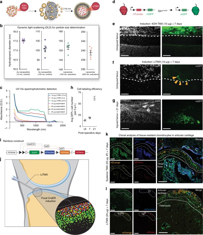
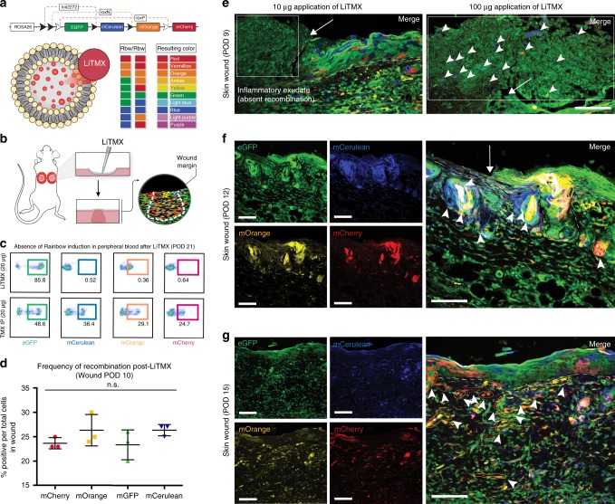
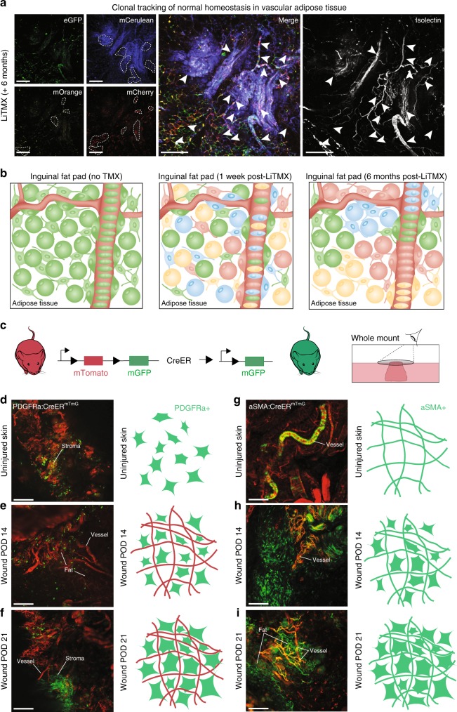
Targeted genetic dissection of tissues to identify precise cell populations has vast biological and therapeutic applications. Here we develop an approach, through the packaging and delivery of 4-hydroxytamoxifen liposomes (LiTMX), that enables localized induction of CreERT2 recombinase in mice. Our method permits precise, in vivo, tissue-specific clonal analysis with both spatial and temporal control. This technology is effective using mice with both specific and ubiquitous Cre drivers in a variety of tissue types, under conditions of homeostasis and post-injury repair, and is highly efficient for lineage tracing and genetic analysis. This methodology is directly and immediately applicable to the developmental biology, stem cell biology and regenerative medicine, and cancer biology fields.


Conflict of interest statement

The authors declare no competing interests.

Figures

Fig. 1
Fig. 1
Generation of LiTMX for targeted tissue labeling. a Schematic of LiTMX generation and extrusion to 100 nm diameter. b Liposomal stability as determined by DLS. Hydrodynamic diameter after filtered extrusion of gold (Au) control nanoparticles at 10-nm core size (purple) and 100-nm core size (blue), and for liposomes at 100 nm (green) and 200 nm extrusion (orange). Experiment conducted with n = 5 replicates per condition. c Stability of LiTMX at 24 h (green), maintained at 6 (red) and 12 weeks (blue) after production, using UV-Vis spectrophometric detection. Following production of LiTMX at various concentrations (10 μg—purple, 20 μg—orange, 30 μg—black, 40 μg—gray), the supernatants show uniformly minimal tamoxifen content demonstrating near complete uptake by the liposomes. d Cre-driven recombination resulting in GFP expression in the bone marrow compartment. e Local application of non-liposomal 4-hydroxytamoxifen (10 μg) to the femoral periosteum (white dotted lines indicate the superficial periosteum and femur cortical bone border, left panels—white indicates DAPI, right panels—green indicates GFP). f In comparison to (e), with application of LiTMX at the same dose (individual GFP+ cells indicated by yellow arrowheads). g Twenty-one days after limb injury, clonal expansion of labeled cells involved in healing occurs along the bone periosteal surface. h The average size of GFP+ clones from g increases with time post-injury (UI = uninjured, n = 5 replicates per timepoint, error bars indicate standard deviation, SD). i Schematic of the Rainbow mouse construct. j Schematic of CreER-driven Rainbow construct induction by LiTMX localized to the articular cartilage. Inset shows whole mount of Rainbow labeled tissue. k Systemically induced (20 μg, IP) resident chondrocytes of the articular cartilage. (Individual channels at left, merged at right, dotted white line indicates articular cartilage, solid white line indicates tibial bone edge, and solid colored lines indicate individual cell clones). l Locally labeled (20 μg) articular cartilage resident chondrocytes by LiTMX in k. Experiments conducted with n = 3 replicates per timepoint (where applicable) per condition (unless otherwise indicated), error bars denote standard error of the mean (s.e.m., unless otherwise indicated), scale bars represent 200 μm (unless otherwise indicated)
Fig. 2
Fig. 2
Application of LiTMX labeled cells in wound healing. a Schematic of the Rainbow reporter mouse construct showing random Cre-mediated recombination after LiTMX induction and resultant cellular color code. Mice homozygous for the Rainbow construct harbor the potential for 10 different colors after recombination. b Rainbow cell labeling in wound healing is achieved via local application of LiTMX using a splinted, dorsal, full-thickness excisional wound model. c Fluorescent activated cell sorting (FACS) analysis of peripheral blood from mice treated with LiTMX (20 μg) (top panels) versus systemic tamoxifen administrated IP (20 μg) (bottom panels). d Randomization of color-coding in activated Rainbow wound tissue shows equal distribution of each fluorescent-labeled cell color (mCherry, mOrange, eGFP, mCerulean) at POD 10 after wounding. e With topical application of LiTMX, in vivo clonal analysis of cutaneous wound healing is visualized at POD 9 on confocal imaging of 2D sectioned specimens (left panel: ×20, white arrow indicates wound edge, white dotted line indicates dermo-epidermal junction of skin, white inset box indicates systemic inflammatory infiltrate with absent recombination; right panel: ×40, close up of area indicated by white arrow in left panel, arrowheads indicate increased incidence of fluorescent cell labeling with increased LiTMX dose). f Labeled clone size increases at POD 12. (Individual channels at left, merged at right, white arrow indicates wound edge, white dotted line indicates dermo-epidermal junction of skin, solid colored lines highlight individual colored cell clones). g Clone size continues to increase at time of wound closure, POD 15. Experiments conducted with n = 3 replicates per timepoint (where applicable) per condition (unless otherwise indicated), 2 dorsal wounds per mouse, error bars denote standard error of the mean (s.e.m.), scale bars represent 200 μm (unless otherwise indicated)
Fig. 3
Fig. 3
LiTMX application in Rainbow mice for clonal analysis in wound healing. a Schematic of Cre-mediated recombination and resultant cellular color code change in Rainbow mice. b Schematic shows splinted, full-thickness excisional skin wounds with local application of LiTMX. c Healing cutaneous wounds are excised, whole mounted and 3D confocal imaging is conducted. d A ×10 magnification of H&E staining of a dorsal wound on cross-section (left), white solid box magnified at ×20 reveals linear migration of cells involved in wound healing (right) (black arrows indicate direction of cell migration). e Whole-mount wound at POD 15 visualized using Imaris software shows linear migration from the wound edge towards the center of fluorescent-labeled clones of cells involved in wound healing. f The average number of cells per clone increases with time post-wounding (data quantified from gi, n = 5 replicates per timepoint, error bars indicate SD). g After local LiTMX dosing at time of cutaneous wounding in Rainbow mice, confocal imaging of whole-mount specimens shows clonal outgrowth of tissue-resident (non-circulating) wound-responsive cells at POD 10. (eG = eGFP, mCe = mCerulean, mOr = mOrange, and mCh = mCherry, individual channels at left, merged images at right). h Clone size increases at POD 14 (white staining, inset, indicates isolectin staining to highlight vasculature). i Clone size continues to increase during remodeling after the wound is healed, at POD 21. Experiments conducted with n = 3 replicates per timepoint (where applicable) per condition (unless otherwise indicated), 2 dorsal wounds per mouse, scale bars represent 200 μm (unless otherwise indicated)
Fig. 4
Fig. 4
LiTMX application for tracing vasculogenic lineages. a Clonal tracking of adipose tissue involved with vasculature in the inguinal fat pad in Rainbow mice at 6 months after LiTMX application (left panel shows split channels, middle panel—merged). Isolectin stains cells engaged with vasculature (right panel). White dotted lines (left) and arrowheads (middle and right) mark clonal cell populations. b Schematic illustrating the fat pad prior to LiTMX application (left panel), 1 week after application with LiTMX-mediated recombination labeling individual cells (middle panel), and the expansion of clonal populations in uninjured fat pad tissue 6 months after induction (right panel). ci Schematic of experimental application of LiTMX to uninjured skin in PDGFRα-CreERT2::Rosa26-mTmG (left panels, df) and αSMA-CreERT2::Rosa26-mTmG (right panels, gi) mice: c Schematic of the mTmG mouse construct. d, g LiTMX labeling results in highly-specific localized activation of cells engaged with fibrous tissue and vasculature, respectively, in uninjured skin at homeostasis. (White labeling of wound structures as indicated, confocal images at left in each panel, schematics at right highlight induced cellular phenotypes based on the Cre drivers used). e, h At POD 14 after cutaneous wounding and LiTMX induction, the PDGFRα-CreERT2::Rosa26-mTmG construct highlights activated fibrogenic cells involved in wound healing, while the αSMA-CreERT2::Rosa26-mTmG construct highlights stromal cells associated with vasculature. f, i At POD 21 after cutaneous wounding, the cells seen at POD 14 (e, h), clonally expand. Experiments conducted with n = 3 replicates per timepoint (where applicable) per condition (unless otherwise indicated), 2 dorsal wounds per mouse (where applicable), scale bars represent 200 μm (unless otherwise indicated)

References

    1. Sauer B, Henderson N. Site-specific DNA recombination in mammalian cells by the Cre recombinase of bacteriophage P1. Proc. Natl Acad. Sci. USA. 1988;85:5166–5170. doi: 10.1073/pnas.85.14.5166. - DOI - PMC - PubMed
    1. Akagi K, et al. Cre-mediated somatic site-specific recombination in mice. Nucleic Acids Res. 1997;25:1766–1773. doi: 10.1093/nar/25.9.1766. - DOI - PMC - PubMed
    1. Feil S, Valtcheva N, Feil R. Inducible Cre mice. Methods Mol. Biol. 2009;530:343–363. doi: 10.1007/978-1-59745-471-1_18. - DOI - PubMed
    1. Sinha DK, et al. Photoactivation of the CreER T2 recombinase for conditional site-specific recombination with high spatiotemporal resolution. Zebrafish. 2010;7:199–204. doi: 10.1089/zeb.2009.0632. - DOI - PubMed
    1. Bhatia A, Kumar R, Katare OP. Tamoxifen in topical liposomes: development, characterization and in-vitro evaluation. J. Pharm. Pharm. Sci. 2004;7:252–259. - PubMed

Publication types

MeSH terms

LinkOut - more resources