Review
. 2018 Jul 13:9:890.
doi: 10.3389/fphys.2018.00890. eCollection 2018.

Nanotherapeutics for Treatment of Pulmonary Arterial Hypertension

Affiliations
Review

Nanotherapeutics for Treatment of Pulmonary Arterial Hypertension

Victor Segura-Ibarra et al. Front Physiol. .

Abstract

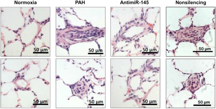
Pulmonary arterial hypertension (PAH) is a devastating and fatal chronic lung disease. While current pharmacotherapies have improved patient quality of life, PAH drugs suffer from limitations in the form of short-term pharmacokinetics, instability, and poor organ specificity. Traditionally, nanotechnology-based delivery strategies have proven advantageous at increasing both circulation lifetimes of chemotherapeutics and accumulation in tumors due to enhanced permeability through fenestrated vasculature. Importantly, increased nanoparticle (NP) accumulation in diseased tissues has been observed pre-clinically in pathologies characterized by endothelial dysfunction and remodeled vasculature, including myocardial infarction and heart failure. Recently, this phenomenon has also been observed in preclinical models of PAH, leading to the exploration of NP-based drug delivery as a therapeutic modality in PAH. Herein, we discussed the advantages of NPs for efficacious treatment of PAH, including heightened therapeutic delivery to diseased lungs for increased drug bioavailability, as well as highlighted innovative nanotherapeutic approaches for PAH.

Keywords: chronic lung disease; drug delivery; nanomedicine; nanoparticles; pulmonary arterial hypertension.


Figures

FIGURE 1
FIGURE 1
Nanoparticle platforms explored in PAH drug and gene delivery. (A) Liposomes are comprised of a lipid bilayer, with an aqueous core ideal for encapsulation of water-soluble drugs (red). (B) Polymer micelles are comprised of amphiphilic block copolymers that self-assemble in water to form a hydrophilic corona and a hydrophobic core for encapsulation of lipophilic drugs (violet). (C) Solid polymer particles have drug dissolved or embedded within a polymer matrix.
FIGURE 2
FIGURE 2
Schematic representation of endothelial dysfunction in PAH and a proposed mechanism of NP extravasation into pulmonary vasculature. While healthy pulmonary vasculature is a semipermeable membrane barrier, the vasculature in lungs undergoing PAH exhibits endothelial dysfunction arising from a chronic inflammatory state and hypoxia, leading to fenestrations (openings) in the endothelium and a hyperpermeable state. Such permeability may be exploited by NPs to passively extravasate and accumulate in lungs undergoing PAH. Figure adapted from Segura-Ibarra et al. (2017), reproduced with permission courtesy of Elsevier.
FIGURE 3
FIGURE 3
NP accumulation in PAH lung vasculature. (A) LC/MS analysis of rapamycin (RAP) concentration in lung tissues 2 and 24 h after a single administration of 15 mg/kg of RAP, either as a free drug formulation (RAP FD) or nanoparticle form (RAP NP) in healthy and MCT-induced model of PH in rats (PH). Results represent mean ± SEM (∗∗∗∗P < 0.0001). (B) Confocal imaging depicting fluorescently loaded NPs in diseased lungs. CD31 positive endothelial cells appear in yellow, NPs are green, while DAPI appears as blue. The scale bar represents 25 μm. (C) Surface intensity plot of the image from panel B representing NP signal. Figure adapted from Segura-Ibarra et al. (2017), reproduced with permission courtesy of Elsevier.
FIGURE 4
FIGURE 4
Effects on survival and RV systolic pressures following pitavastatin-NP delivery. (A) Kaplan–Meier curve depicting survival of MCT-induced PAH rats following a single intratracheal administration of Pitavastatin-NP compared to controls consisting of intratracheal delivery of PBS, free pitavastatin, and fluorescein isothiocyanate (FITC)-NPs. (B) Effects of pitavastatin-NPs on RV systolic pressure (expressed as mmHg). P < 0.01 vs. untreated control. Figure adapted from Chen et al. (2011), reproduced with permission courtesy of Wolters Kluwer Health, Inc.
FIGURE 5
FIGURE 5
Rapamycin NPs prevented pulmonary arteriole hypertrophy in PAH and did not lead to an increase in inflammatory cytokines. (A) Verhoeff–Van Gieson (VVG) stain of pulmonary arterioles from MCT-induced model of PAH in rats treated with free rapamycin (RAP FD), NP vehicle (Vehicle), and RAP NPs. Scale bars represent 50 μm. (B) Quantification of the relative wall thickness among treated groups in (A). Results shown as mean ± SEM (∗∗∗∗P < 0.0001). Serum levels of inflammatory cytokines TNF-α (C) and IL-1β (D) measured after the course of treatment. Results represent mean ± SEM values (∗∗P < 0.01, P < 0.05). Figure adapted from Segura-Ibarra et al. (2017), reproduced with permission courtesy of Elsevier.
FIGURE 6
FIGURE 6
Effects of anti-miR-145 loaded liposomes on arteriole hyperplasia in an MCT-induced model of PAH. Hematoxylin and Eosin (H&E) stained histological sections depicting pulmonary arterioles following anti-miR-145 liposome treatment of rats with Sugen/Hypoxia induced PAH. Results highlight anti-miR-145 liposome treatment compared to controls consisting of healthy controls (normoxia), rats undergoing PAH (PAH), and liposomes containing non-silencing control oligonucleotide (non-silencing). Scale bar represents 50 μm. Figure adapted from McLendon et al. (2015), reproduced with permission courtesy of Elsevier.

References

    1. Acharya S., Sahoo S. K. (2011). PLGA nanoparticles containing various anticancer agents and tumour delivery by EPR effect. Adv. Drug Deliv. Rev. 63 170–183. 10.1016/j.addr.2010.10.008 - DOI - PubMed
    1. Akagi S., Nakamura K., Matsubara H., Kondo M., Miura D., Matoba T., et al. (2016). Intratracheal administration of prostacyclin analogue-incorporated nanoparticles ameliorates the development of monocrotaline and sugen-hypoxia-induced pulmonary arterial hypertension. J. Cardiovasc. Pharmacol. 67 290–298. 10.1097/FJC.0000000000000352 - DOI - PMC - PubMed
    1. Akagi S., Nakamura K., Miura D., Saito Y., Matsubara H., Ogawa A., et al. (2015). Delivery of imatinib-incorporated nanoparticles into lungs suppresses the development of monocrotaline-induced pulmonary arterial hypertension. Int. Heart J. 56 354–359. 10.1536/ihj.14-338 - DOI - PubMed
    1. Anderson R., Franch A., Castell M., Perez-Cano F. J., Brauer R., Pohlers D., et al. (2010). Liposomal encapsulation enhances and prolongs the anti-inflammatory effects of water-soluble dexamethasone phosphate in experimental adjuvant arthritis. Arthritis Res. Ther. 12:R147. 10.1186/ar3089 - DOI - PMC - PubMed
    1. Aversa M., Porter S., Granton J. (2015). Comparative safety and tolerability of endothelin receptor antagonists in pulmonary arterial hypertension. Drug Saf. 38 419–435. 10.1007/s40264-015-0275-y - DOI - PubMed