. 2018 Jun 27:4:3.
doi: 10.1038/s41420-018-0068-z. eCollection 2018.

Guanylate binding proteins facilitate caspase-11-dependent pyroptosis in response to type 3 secretion system-negative Pseudomonas aeruginosa

Affiliations

Guanylate binding proteins facilitate caspase-11-dependent pyroptosis in response to type 3 secretion system-negative Pseudomonas aeruginosa

Arjun Balakrishnan et al. Cell Death Discov. .

Erratum in

Abstract

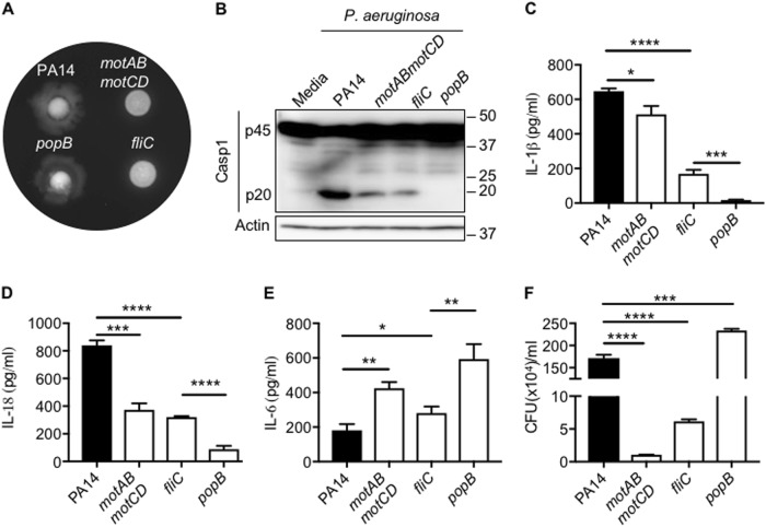
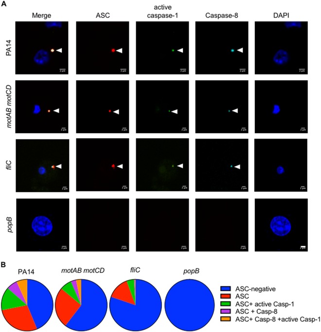
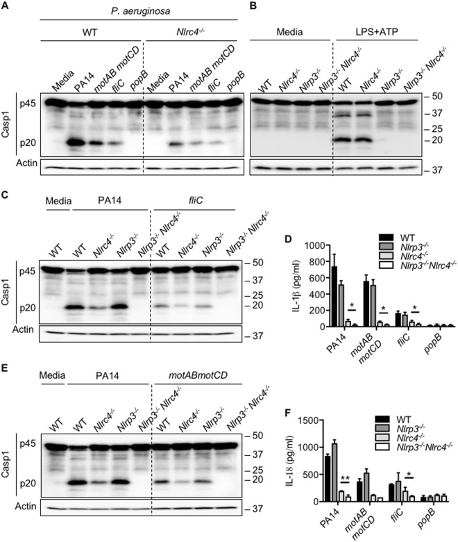
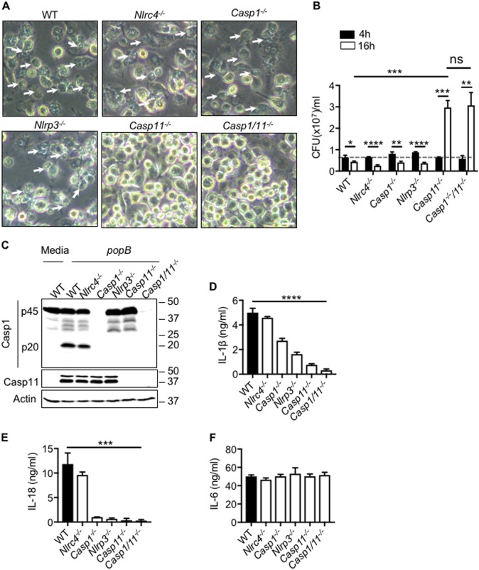
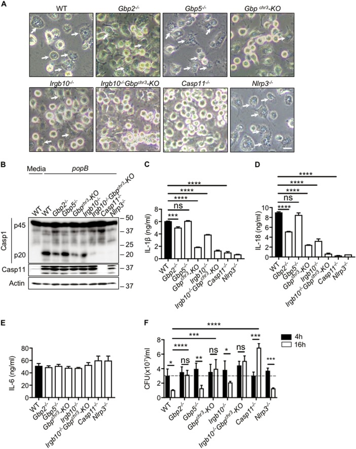
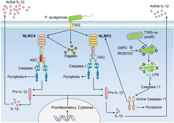
Detection of bacterial ligands is a pre-requisite for inflammasome activation. During Pseudomonas aeruginosa infection, flagellin which is secreted through the T3SS is detected by the NLRC4 inflammasome. Activation of the NLRC4 inflammasome is believed to contribute to high IL-1β production and pathogenicity in cystic fibrosis patients with chronic P. aeruginosa infection. Interestingly, the majority of P. aeruginosa isolated from cystic fibrosis patients with chronic airway infection are non-motile and T3SS-negative, suggesting that yet un-characterized inflammasome pathways regulate IL-1β production in cystic fibrosis patients. Here we demonstrate the role of guanylate-binding proteins (GBPs) in regulating bacterial proliferation and inflammasome activation in response to T3SS-negative P. aeruginosa. Bacterial ligands liberated by the action of GBP2 and IRGB10 activate caspase-11 and regulate non-canonical NLRP3 inflammasome activation and IL-1β release. Overall, our results reveal the role of caspase-11 in inhibiting bacterial proliferation and promoting IL-1β secretion during T3SS-negative P. aeruginosa infection. This study suggests that non canonical inflammasomes might have co-evolved to detect Gram-negative bacterial pathogens that have evolved to bypass detection by canonical NLRs.


Conflict of interest statement

The authors declare that they have no conflict of interest.

Figures

Fig. 1
Fig. 1. Non-motile P. aeruginosa activates caspase-1 through T3SS.
a The motility phenotype of strains (PA14, motAB motCD, fliC, popB) confirmed in soft agar. be Caspase-1 activation, IL-1β, IL-18, and IL-6 release in unprimed bone marrow-derived macrophages (BMDMs) infected with indicated strains of P. aeruginosa (MOI 10) for 2 h. f Intracellular bacterial numbers in unprimed BMDMs infected with indicated strains of P. aeruginosa (MOI 1) for 30 min. Data are representative of two (a, f) or three (be) independent experiments. *p < 0.05, **p < 0.01, ***p < 0.001, ****p < 0.0001 (Two-tailed T test)
Fig. 2
Fig. 2. ASC foci formation in macrophages by P. aeruginosa is T3SS dependent.
a Unprimed BMDMs were infected with indicated strains of P. aeruginosa (MOI 10) for 1 h and stained for ASC (red), active caspase-1 (green), caspase-8 (magenta) and DNA (blue). Arrow heads indicate an inflammasome complex. (Scale, 2 μm). b Composition of ASC specks. At least 200 BMDMs infected with indicated strains were counted
Fig. 3
Fig. 3. Pseudomonas aeruginosa T3SS induces NLRP3 inflammasome in the absence of NLRC4 inflammasome.
a Immunoblot analysis of caspase-1 in unprimed WT, Nlrc4/ BMDMs left untreated or infected with indicated strains of P. aeruginosa (MOI 10) for 2 h. b Immunoblot analysis of caspase-1 in unprimed or LPS-primed WT, Nlrc4/, Nlrp3/ and Nlrp3/Nlrc4/ BMDMs stimulated with ATP (LPS + ATP). cf Immunoblot analysis of caspase-1, IL-1β, and IL-18 release in unprimed WT, Nlrc4/, Nlrp3/ and Nlrp3/Nlrc4/ BMDMs left untreated or infected with indicated strains of P. aeruginosa (MOI 10) for 2 h. Data are representative of three independent experiments. *p < 0.05, **p < 0.01 (two-tailed T test)
Fig. 4
Fig. 4. Caspase-11 mediated pyroptosis restrict proliferation of T3SS mutant in macrophages.
a Microscopic analysis of cell death in unprimed WT, Nlrc4/, Caspase1/, Nlrp3/,Caspase11/ and Caspase1/ Caspase11/ BMDMs infected with popB (MOI 10) for 16 h. (Scale, 15 μm). Arrow heads indicate pyroptotic cells. b Bacterial CFU in unprimed WT, Nlrc4/, Caspase1/, Nlrp3/, Caspase11/ and Caspase1/ Caspase11/ BMDMs infected with popB MOI10, 4 h and 16 h. c-f Immunoblot analysis of caspase-1, IL-1β, IL-18, and IL-6 release in unprimed WT, Nlrc4/, Caspase1/, Nlrp3/, Caspase11/, and Caspase1/ Caspase11/ BMDMs left untreated or infected with popB for 16 h. Data are representative of two (b) or three (a,cf) independent experiments. ns not significant, *p < 0.05, **p < 0.01, ***p < 0.001, ****p < 0.0001 (two-tailed T test)
Fig. 5
Fig. 5. GBP2 and IRGB10 regulates caspase-11 mediated non-canonical NLRP3 activation and pyroptosis.
a Microscopic analysis of cell death in unprimed WT, Gbp2/, Gbp5/, Gbpchr3-KO,Irgb10/, Irgb10/ Gbpchr3-KO, Caspase11/ and Nlrp3/ BMDMs infected with popB (MOI 10) for 16 h. (Scale, 15 μm). Arrow heads indicate pyroptotic cells. be Immunoblot analysis of caspase-1, IL-1β, IL-18, and IL-6 release in WT, Gbp2/, Gbp5/, Gbpchr3-KO, Irgb10/, Irgb10/ Gbpchr3-KO, Caspase11/ and Nlrp3/ BMDMs infected with popB (MOI10). f Bacterial CFU in unprimed WT, Gbp2/, Gbp5/, Gbpchr3-KO, Irgb10/, Irgb10/ Gbpchr3-KO, Caspase11/ and Nlrp3/ BMDMs infected with popB (MOI10), 4 h and 16 h. Data are representative of three independent experiments. ns-not significant, *p < 0.05, **p < 0.01, ***p < 0.001,****p < 0.0001 (two-tailed T test)
Fig. 6
Fig. 6. Model depicting inflammasome activation by WT and T3SS-negative P.aeruginosa during acute and chronic infection in macrophages.
During acute infection (2 h), bacterial T3SS itself and flagellin secreted through T3SS activates NLRC4 inflammasome. T3SS also activates NLRP3 inflammasome by yet uncharacterized mechanisms. T3SS-negative P.aeruginosa escapes detection by both NLRP3 and NLRC4 inflammasomes and proliferates inside cytoplasm. During chronic infection (16 h), high numbers of T3SS-negative P.aeruginosa is detected by GBP2 and IRGB10. Bacterial lysis by action of GBPs release ligands that lead to activation of caspase-11. Caspase-11 inhibits the proliferation of bacteria and also activates NLRP3 inflammasome which leads to secretion of inflammatory cytokine IL-1β

References

    1. Franchi L, et al. Critical role for Ipaf in Pseudomonas aeruginosa-induced caspase-1 activation. Eur. J. Immunol. 2007;3711:3030–3039. doi: 10.1002/eji.200737532. - DOI - PubMed
    1. Bonfield TL, et al. Inflammatory cytokines in cystic fibrosis lungs. Am. J. Respir. Crit. Care. Med. 1995;1526(Pt 1):2111–2118. doi: 10.1164/ajrccm.152.6.8520783. - DOI - PubMed
    1. Konstan MW, Davis PB. Pharmacological approaches for the discovery and development of new anti-inflammatory agents for the treatment of cystic fibrosis. Adv. Drug Deliv. Rev. 2002;5411:1409–1423. doi: 10.1016/S0169-409X(02)00146-1. - DOI - PubMed
    1. Cohen TS, Prince AS. Activation of inflammasome signaling mediates pathology of acute P. aeruginosa pneumonia. J. Clin. Invest. 2013;1234:1630–1637. doi: 10.1172/JCI66142. - DOI - PMC - PubMed
    1. Sharma D, Kanneganti TD. The cell biology of inflammasomes: mechanisms of inflammasome activation and regulation. J. Cell. Biol. 2016;2136:617–629. doi: 10.1083/jcb.201602089. - DOI - PMC - PubMed