

Otogenic brain abscesses: A systematic review
- PMID: 30062135
- PMCID: PMC6057212
- DOI: 10.1002/lio2.150
Otogenic brain abscesses: A systematic review
Abstract
Objective: Otogenic brain abscesses are one of the most significant life-threatening complications of otologic infections. Given their low prevalence, otogenic brain abscesses require a high index of suspicion for diagnosis. In this systematic review, we aim to provide an analysis of otogenic brain abscesses and describe common clinical signs and symptoms, bacteriology, location, treatment options, morbidity, and mortality.
Data sources: PubMed, Cochrane CENTRAL database, Google Scholar, and Scopus.
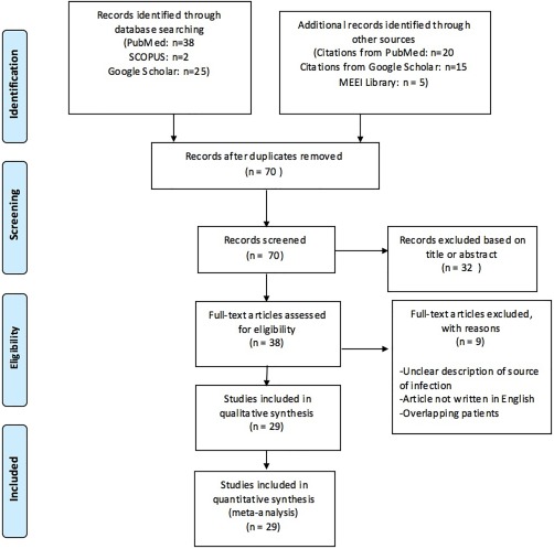
Methods: A systematic review of literature was performed using the Preferred Reporting Items for Systematic Reviews and Meta-analyses recommendations. Variables assessed included clinical signs and symptoms, bacteriology, location, treatment, morbidity, and mortality.
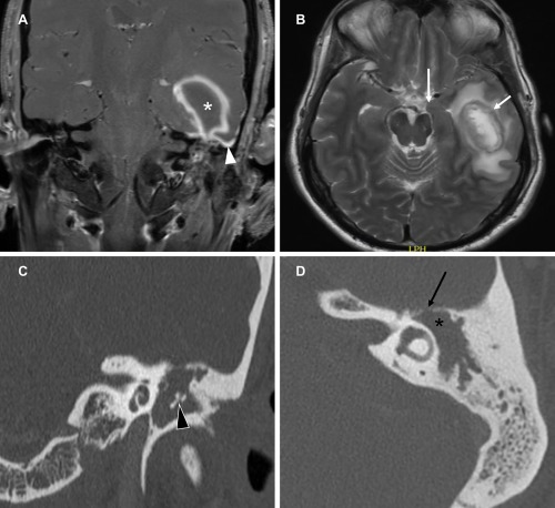
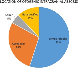
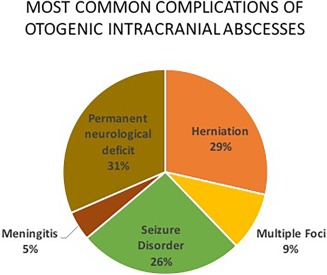
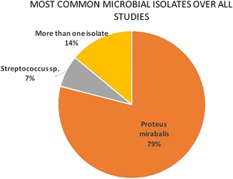
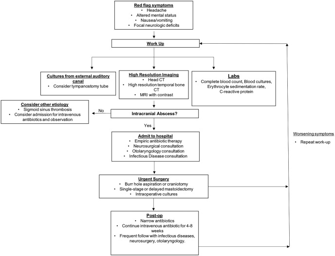
Results: Twenty-nine studies met inclusion and exclusion criteria, corresponding to a total of 1307 otogenic abscess cases for review. Fifty-five percent of abscesses were found in the temporal lobe and 28% in the cerebellum. Most patients (88.3%) had a history of suppurative chronic otitis media. The most common symptoms were headache, altered mental status, papilledema, and meningeal irritation. Fever, nausea, and vomiting affected about 40% of patients. The most commonly cultured bacterial species was Proteus mirabilis. In addition to antibiotics, most otogenic brain abscesses were treated by burr hole aspiration. Average mortality following advent of computed tomography was 8.11%.
Conclusion: Although rare, otogenic brain abscesses may occur as a complication of suppurative otitis media and require a high index of suspicion. Appropriate imaging studies and multidisciplinary expertise are crucial in the diagnosis and management.
Level of evidence: 4.
Keywords: Brain abscess; computed tomography; magnetic resonance imaging; otologic infection.
Figures
References
-
- Wintermeyer SM, Nahata MC. Chronic suppurative otitis media. Ann Pharmacother 1994;28:1089–1099. - PubMed
-
- Harkness P, Topham J. Classification of otitis media. Laryngoscope 1998;108:1539–1543. - PubMed
-
- Yorgancilar E, Akkus Z, Gun R, et al. Temporal bone erosion in patients with chronic suppurative otitis media. B‐ENT 2013;9:17–22. - PubMed
LinkOut - more resources
Full Text Sources
Other Literature Sources
Research Materials
Miscellaneous