. 2017 Oct 30;2(5):591-600.
doi: 10.1016/j.jacbts.2017.06.004. eCollection 2017 Oct.

Apolipoprotein E Deficiency Increases Remnant Lipoproteins and Accelerates Progressive Atherosclerosis, But Not Xanthoma Formation, in Gene-Modified Minipigs

Affiliations

Apolipoprotein E Deficiency Increases Remnant Lipoproteins and Accelerates Progressive Atherosclerosis, But Not Xanthoma Formation, in Gene-Modified Minipigs

Jeong Shim et al. JACC Basic Transl Sci. .

Abstract

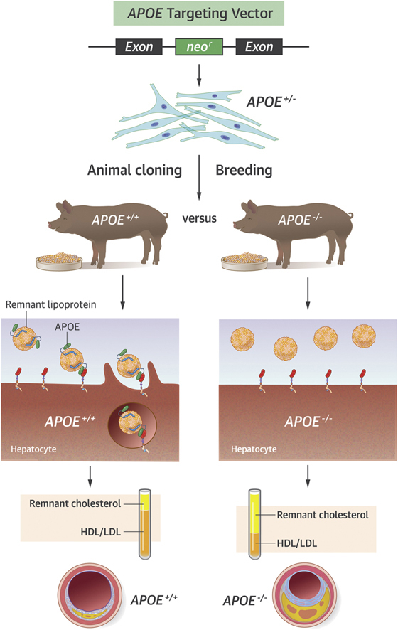
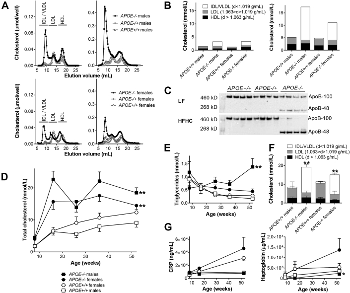
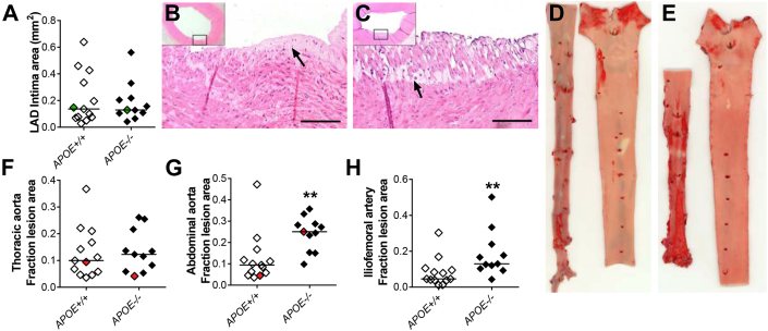
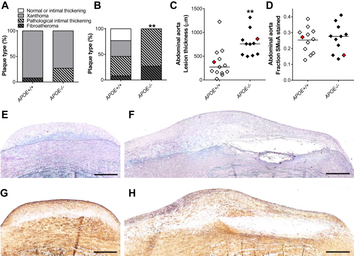
Deficiency of apolipoprotein E (APOE) causes familial dysbetalipoproteinemia in humans resulting in a higher risk of atherosclerotic disease. In mice, APOE deficiency results in a severe atherosclerosis phenotype, but it is unknown to what extent this is unique to mice. In this study, APOE was targeted in Yucatan minipigs. APOE-/- minipigs displayed increased plasma cholesterol and accumulation of apolipoprotein B-48-containing chylomicron remnants on low-fat diet, which was significantly accentuated upon feeding a high-fat, high-cholesterol diet. APOE-/- minipigs displayed accelerated progressive atherosclerosis but not xanthoma formation. This indicates that remnant lipoproteinemia does not induce early lesions but is atherogenic in pre-existing atherosclerosis.

Keywords: APOB, apolipoprotein B; APOE, apolipoprotein E; HFHC, high-fat high-cholesterol; IDL, intermediate-density lipoprotein; LAD, left anterior descending (coronary artery); LDL, low-density lipoprotein; LDLR, low-density lipoprotein receptor; LF, low-fat; Neo, neomycin; SMC, smooth muscle cell; VLDL, very-low-density lipoprotein; apolipoprotein E; atherosclerosis; cDNA, complementary DNA; pig; rAAV, recombinant adeno-associated virus; remnant cholesterol dysbetalipoproteinemia.


Figures

None
Graphical abstract
Figure 1
Figure 1
APOE Gene Targeting (A) Schematic representation of the endogenous APOE locus, the gene targeting vector and the targeted APOE locus. The exons of the endogenous porcine APOE gene are shown as black boxes. Expression of the neor cDNA cassette is driven by a PGK promoter, whereas expression of the zeor cDNA cassette used for bacterial selection is driven by an EM7 promoter. A region of 295 bp in APOE, comprising the entire exon 3 and its flanking regions, is upon successful gene targeting replaced by the PGK-neor-EM7-zeor cassette resulting in a targeted DNA-fragment of 5,530 bp when XmnI-digested genomic DNA is hybridized with the APOE- or neor-specific probe. Positions of the PCR screening primer pairs F1/R1 and F2/R2 are indicated by small horizontal arrows. XmnI restriction sites are indicated by vertical arrows and Southern blot probes (APOEprobe and neorprobe) are illustrated as black horizontal bars. (B) Representative Southern blot of genomic DNA isolated from cloned APOE+/− Yucatan minipigs. XmnI-digested genomic DNA was electrophoresed, blotted on to a nitrocellulose membrane, and hybridized with the APOE probe resulting in a 5.5 kb and a 11.9 kb band representing the targeted and the wild-type allele, respectively (upper panel). The genomic DNA was also hybridized with the neor probe, detecting the neor cassette, yielding only the expected targeted 5.5 kb band (lower panel). Lanes 1-6: Genomic DNA from representative cloned APOE+/− Yucatan minipigs. Lane 7: Yucatan APOE+/+ control minipig. (C) Reverse transcriptase PCR. Total RNA was isolated from liver tissue and used for first strand cDNA synthesis. RT-PCR was performed using primers specific for the APOE exon 3 region or the porcine β-actin gene ACTB. PGK = phosphoglycerate kinase; PCR = polymerase chain reaction; RT-PCR = reverse transcriptase polymerase chain reaction.
Figure 2
Figure 2
Characterization of Cholesterol Profiles and Inflammatory Markers (A) Size-exclusion chromatography on plasma pools of cholesterol in APOE+/+, APOE+/−, and APOE−/− Yucatan male and female minipigs on low-fat (LF) diet (left column) and after 8 weeks on high-fat high-cholesterol (HFHC) diet (right column). (B) Ultracentrifugation characterization of cholesterol on plasma pools from APOE+/+ and APOE−/− Yucatan minipigs on LF diet (left) and after 8 weeks on HFHC diet (right). (C) Western blot of APOB in plasma from APOE+/+, APOE+/−, and APOE−/− Yucatan minipigs on LF diet (upper) and on HFHC diet (lower). (D) Enzymatic measurement of total cholesterol at 8 (on LF diet), 16, 26, 36, and 52 (all on HFHC diet) weeks of age. **p = 0.0022 for APOE+/+ male versus APOE−/− male minipigs and p = 0.0051 for APOE+/+ female versus APOE−/− female minipigs. (E) Enzymatic measurement of triglycerides at 8 (on LF diet), 16, 26, 36, and 52 (all on HFHC diet) weeks of age. **p = 0.0087 for APOE+/+ male versus APOE−/− male minipigs. (F) Ultracentrifugation characterization of cholesterol in plasma from APOE+/+, APOE+/−, and APOE−/− Yucatan minipigs on HFHC diet at 52 weeks of age. **p = 0.0022 for IDL/VLDL between APOE+/+ male and APOE−/− male minipigs and p = 0.0025 for IDL/VLDL between APOE+/+ female and APOE−/− female minipigs. p = 0.0303 for HDL and p = 0.0025 for LDL between APOE+/+ female and APOE−/− female minipigs. (G) C-reactive protein (CRP) (left) and haptoglobin (right) levels in APOE+/+ and APOE−/− Yucatan minipigs at 8, 16, and 52 weeks of age. *p = 0.0411 for APOE+/+ male versus APOE−/− male minipigs. (D to G)APOE+/+ males n = 6, APOE+/+ females n = 7, and APOE−/− males n = 6, and APOE−/− females n = 5. Error bars indicate SEM. HDL = high-density lipoprotein; IDL = intermediate-density lipoprotein; VLDL = very-low-density lipoprotein.
Figure 3
Figure 3
Lesion Quantification (A) Average cross-sectional intima area in left anterior descending (LAD) coronary artery. (B, C) Representative examples (green dots in A) of lesions in the LAD of an APOE+/+(B) and an APOE−/−(C) Yucatan minipig. Arrows indicate examples of foam cells. Scale bar = 100 μm. Both are examples of xanthomas with intima containing foam cells. (D, E) Representative examples (red dots in F and G) of en face stained aortic lesions from an APOE+/+(D) and an APOE−/−(E) Yucatan minipig. In both, abdominal aorta is depicted left and thoracic aorta depicted right. Red areas (stained with Sudan IV) represent lipid-rich areas of the aortic intima. (F) Surface covered with red-stained area in the thoracic aorta (mainly non-raised lesions). (G) Surface covered with red-stained area in the abdominal aorta (mainly raised lesions). **p = 0.0020. (H) Surface covered with red-stained area in the iliofemoral arteries. **p = 0.0059. In A, F to H: APOE+/+ n = 13 and APOE−/− n = 11. Bars indicate median.
Figure 4
Figure 4
Lesion Characterization (A) Plaque-classification of the most advanced lesion in the LAD coronary artery of each animal. (B) Plaque classification of the most raised lesion in close proximity to the aortic trifurcation. **p = 0.0059. (C) Thickness of the most raised lesion in close proximity to the aortic trifurcation. Bars indicate median. **p = 0.012. (D) Area stained positive for SMαA in the most raised lesion in close proximity to the aortic trifurcation. Bars indicate mean. (E, F) Representative examples (red dots in C and D) of elastin trichrome stained lesions in the most raised lesion in close proximity to the aortic trifurcation of an APOE+/+(E) and an APOE−/−(F) Yucatan minipig. Scale bar = 500 μm. (G, H) Same lesions as in E and F, respectively, immunohistochemically stained for SMαA. (A to D)APOE+/+ n = 13 and APOE−/− n = 11. SMαA = smooth muscle α-actin; other abbreviation as in Figure 3.

References

    1. Plump A.S., Smith J.D., Hayek T. Severe hypercholesterolemia and atherosclerosis in apolipoprotein E-deficient mice created by homologous recombination in ES cells. Cell. 1992;71:343–353. - PubMed
    1. Véniant M.M., Zlot C.H., Walzem R.L. Lipoprotein clearance mechanisms in LDL receptor-deficient “apo-B48- only” and “apo-B100-only” mice. J Clin Invest. 1998;102:1559–1568. - PMC - PubMed
    1. Kim E., Cham C.M., Véniant M.M., Ambroziak P., Young S.G. Dual mechanisms for the low plasma levels of truncated apolipoprotein B proteins in familial hypobetalipoproteinemia. Analysis of a new mouse model with a nonsense mutation in the Apob gene. J Clin Invest. 1998;101:1468–1477. - PMC - PubMed
    1. Getz G.S., Reardon C.A. Apoprotein E as a lipid transport and signaling protein in the blood, liver, and artery wall. J Lipid Res. 2009;50:156–161. - PMC - PubMed
    1. Wu D., Sharan C., Yang H. Apolipoprotein E-deficient lipoproteins induce foam cell formation by downregulation of lysosomal hydrolases in macrophages. J Lipid Res. 2007;48:2571–2578. - PubMed