

Case Reports
doi: 10.1016/j.case.2017.05.005.
eCollection 2017 Oct.
Multimodality Imaging and Pathologic Assessment in an Adult with Endocardial Fibroelastosis
Affiliations
- PMID: 30062275
- PMCID: PMC6058245
- DOI: 10.1016/j.case.2017.05.005
Item in Clipboard
Case Reports
Multimodality Imaging and Pathologic Assessment in an Adult with Endocardial Fibroelastosis
CASE (Phila).
.
Abstract
•The authors report a patient with biopsy-proven adult endocardial fibroelastosis.•Transthoracic echocardiography revealed diffuse coarse endocardial calcifications.•Native CT of the chest revealed LV endocardial calcifications.
Keywords: Adult cardiomyopathy; Adult endocardial fibroelastosis; Multimodality imaging.
Figures

Chest radiograph in the frontal projection showing coarse calcifications over the left heart (black arrow).

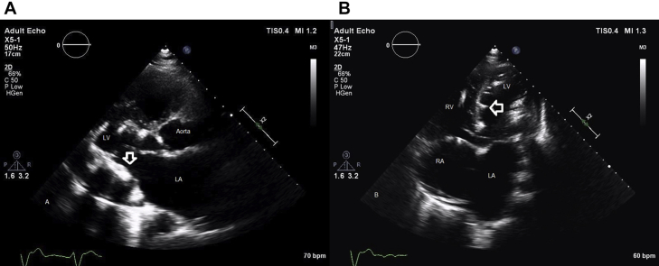
(A,B) Transthoracic echocardiography in the parasternal long-axis and apical four-chamber images showing hyperechoic foci with associated acoustic shadowing of myocardial calcifications (white arrows). LA, Left atrial; LV, left ventricular; RA, right atrial; RV, right ventricular.

Transthoracic echocardiography in the parasternal apical four-chamber images showing central jet of tricuspid regurgitation (white arrow).

(A-C) Four-chamber, short-axis, and two-chamber reformatted images from chest computed tomography without contrast showing coarse endocardial calcification with extensive involvement of the mitral annulus (black arrows). LA, Left atrial; LV, left ventricular; RA, right atrial; RV, right ventricular.

Endomyocardial biopsy. (A) Light micrograph of the subendocardium of a biopsy piece showing wavy eosinophilic material corresponding to fibrous tissue, which thickens the endocardium and is birefringent. This tissue also infiltrates the subjacent myocardium. The myocytes show mild hypertrophy with diameters ranging from 30 to 40 μm. There are no inflammatory infiltrates. Hematoxylin and Eosin stain, 400×. (B) The same field of biopsy shown in (A) distinctly demonstrates elastic fibers (black) interspersed between the dense fibrous tissue (yellow) in the areas of endocardial thickening, thus demonstrating endocardial fibroelastosis. Movat pentachrome stain, 400×.
References
-
- Seki A., Patel S., Ashraf S., Perens G., Fishbein M.C. Primary endocardial fibroelastosis: an underappreciated cause of cardiomyopathy in children. Cardiovasc Pathol. 2013;22:345–350. - PubMed
-
- Ni J., Bowles N.E., Kim Y.H., Demmler G., Kearney D., Bricker J.T. Viral infection of the myocardium in endocardial fibroelastosis. Molecular evidence for the role of mumps virus as an etiologic agent. Circulation. 1997;95:133–139. - PubMed
-
- Hitsumoto T., Ikeda S., Matsukage S., Hamada M. Extensive myocardial calcinosis due to Mycobacterium tuberculosis. Eur Heart J. 2016;37:1195. - PubMed
Publication types
LinkOut - more resources
Full Text Sources
Other Literature Sources
