

Case Reports
doi: 10.1016/j.case.2017.06.005.
eCollection 2017 Oct.
Lipomatous Hypertrophy of the Interatrial Septum: A Case Report and Review of the Literature
Affiliations
- PMID: 30062277
- PMCID: PMC6058280
- DOI: 10.1016/j.case.2017.06.005
Item in Clipboard
Case Reports
Lipomatous Hypertrophy of the Interatrial Septum: A Case Report and Review of the Literature
CASE (Phila).
.
No abstract available
Keywords: Imaging modality; Interatrial septum; Lipomatous hypertrophy; Mass.
Figures


(A) TTE, apical four-chamber view. The red arrow indicates the thickened interatrial septum. (B) CMR of the LASH (star) in four-chamber view: end-diastolic frame of a cine image. Note that LASH spares the fossa ovalis. The arrowhead points to LASH anteriorly to the fossa ovalis. LA, Left atrium; LV, left ventricle; RA, right atrium; RV, right ventricle.

TTE, atypical apical four-chamber view. The yellow arrow indicates LASH. LA, Left atrium; LV, left ventricle; RA, right atrium; RV, right ventricle.

TTE, subcostal view. The white arrow represents the hypertrophied interatrial septal thickness. LA, Left atrium; LV, left ventricle; RA, right atrium; RV, right ventricle.
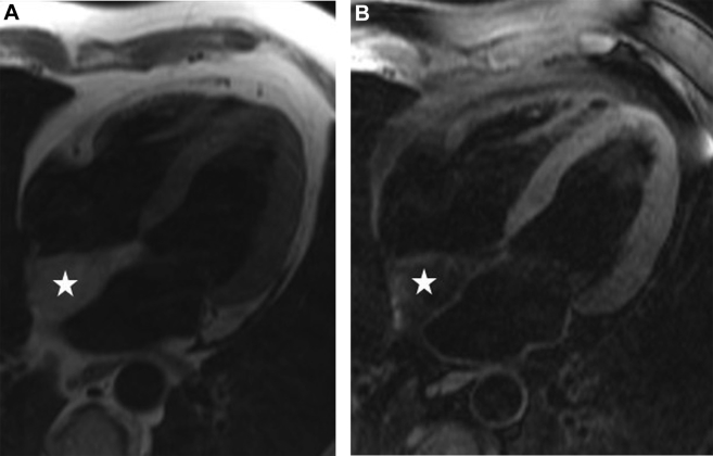
(A) CMR of LASH (star) in the four-chamber view. In T1-weighted images, LASH appears bright. (B) CMR of LASH (star) in the four-chamber view. In T1-weighted images with fat suppression, LASH appears dark. The combination of T1-weighted and T1-weighted images with fat suppression (A,B) is diagnostic for fat.

CMR of LASH (star) in the four-chamber view. In late gadolinium enhancement images, LASH shows mildly increased signal intensity compared with ventricular myocardium.
References
-
- Prior J.T. Lipomatous hypertrophy of cardiac interatrial septum. A lesion resembling hibernoma, lipoblastomatosis and infiltrating lipoma. Arch Pathol. 1964;78:11–15. - PubMed
-
- Heyer C.M., Kagel T., Lemburg S.P., Bauer T.T., Nicolas V. Lipomatous hypertrophy of the interatrial septum: a prospective study of incidence, imaging findings, and clinical symptoms. Chest. 2003;124:2068–2073. - PubMed
-
- Pochis W.T., Saeian K., Sagar K.B. Usefulness of transesophageal echocardiography in diagnosing lipomatous hypertrophy of the atrial septum with comparison to transthoracic echocardiography. Am J Cardiol. 1992;70:396–398. - PubMed
-
- Laura D.M., Donnino R., Kim E.E., Benenstein R., Freedberg R.S., Saric M. Lipomatous atrial septal hypertrophy: a review of its anatomy, pathophysiology, multimodality imaging, and relevance to percutaneous interventions. J Am Soc Echocardiog. 2016;29:717–723. - PubMed
Publication types
LinkOut - more resources
Full Text Sources
Other Literature Sources
