

Case Reports
doi: 10.1016/j.case.2017.10.005.
eCollection 2018 Apr.
An Audaciously Aneurysmal Atrium
Affiliations
- PMID: 30062315
- PMCID: PMC6058409
- DOI: 10.1016/j.case.2017.10.005
Item in Clipboard
Case Reports
An Audaciously Aneurysmal Atrium
CASE (Phila).
.
No abstract available
Keywords: Echocardiography; Imaging; Mitral stenosis; Rheumatic heart disease.
Figures

AP chest radiograph. Anteroposterior chest x-ray revealing alveolar and interstitial opacities in the lung fields, left-sided pneumothorax, and enlarged cardiac silhouette with a cardiothoracic ratio of 0.77.
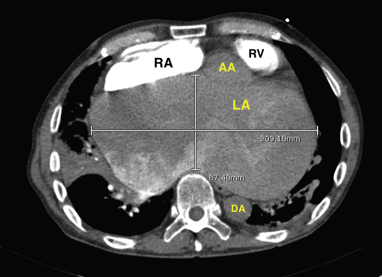
Transverse computed tomographic image of the chest. Computed tomography of the chest depicting the left atrial dimensions including a transverse diameter of 21 cm and an anteroposterior diameter of 8.7 cm. AA, Ascending aorta; DA, descending aorta; LA, left atrium; RA, right atrium; RV, right ventricle.

Coronal computed tomographic image of the chest. Computed tomography of the chest showing superoinferior measurement of the left atrium measured at 17 cm and left-sided pneumothorax (red arrows on the right of the image), and right-sided airspace and interstitial opacities of the lungs (lower left and upper left arrows, respectively). AA, Aortic arch; LA, left atrium; LPA, left pulmonary artery; RPA, right pulmonary artery.
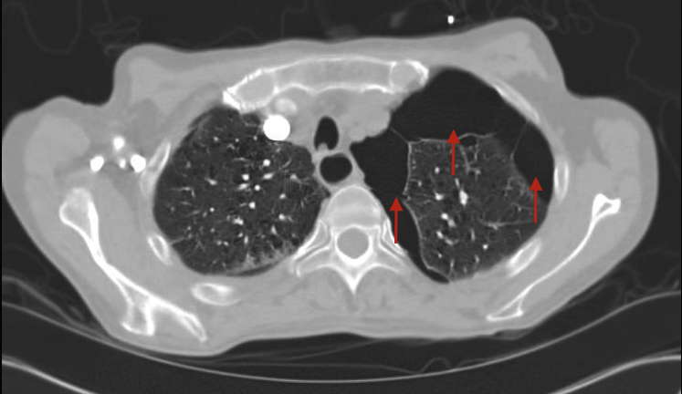
Transverse computed tomographic image of the chest in the lung window. Computed tomography of the chest in the transverse plane revealing multiple pleural blebs (right and left arrows), and a large anterior pneumothorax (center arrow).

Transverse computed tomographic pulmonary angiogram. Computed tomography pulmonary angiographic image showing filling defects in bilateral pulmonary artery branches representing pulmonary emboli (red arrows).
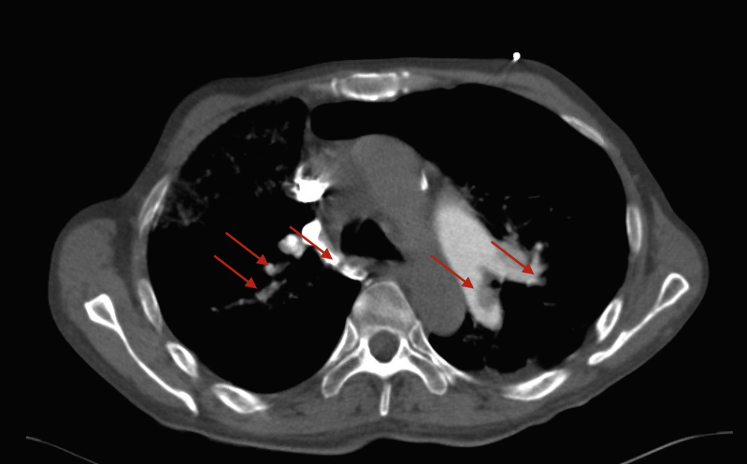
Transverse computed tomographic pulmonary angiogram. Computed tomography pulmonary angiographic image showing filling defects in bilateral pulmonary artery branches representing pulmonary emboli (red arrows).

Transthoracic echocardiogram in parasternal long-axis view. Transthoracic echocardiogram image in parasternal long-axis view depicting a left atrial diameter of 9.2 cm measured in the anteroposterior dimension. DA, Descending aorta; LA, left atrium; LV, left ventricle; LVOT, left ventricular outflow tract; MV, mitral valve.

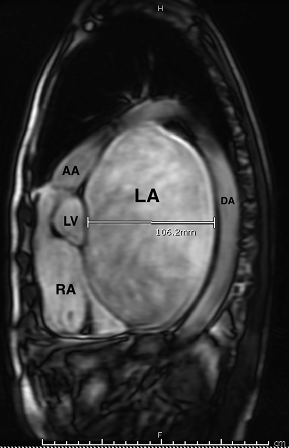
Sagittal image of cardiac magnetic resonance images with depicting anatomy. Sagittal slice of cardiac magnetic resonance imaging revealing the giant left atrium with an anteroposterior diameter of 10.6 cm. AA, Ascending aorta; DA, descending aorta; LA, left atrium; LV, left ventricle; RA, right atrium.

Cardiac magnetic resonance images of left atrial thrombus burden. Cardiac magnetic resonance imaging in the sagittal plane revealing the large burden of left atrial thrombus and microthrombi (white arrows).
References
-
- Piccoli G.P., Massini C., Di Eusanio G., Ballerini L., Iacobone G., Soro A. Giant left atrium and mitral valve disease: early and late results of surgical treatment in 40 cases. J Cardiovasc Surg. 1984;86:595–601. - PubMed
-
- Hurst J.W. Memories of patients with a giant left atrium. Circulation. 2001;104:2630–2631. - PubMed
-
- Oh J.K. Echocardiographic evaluation of morphological and hemodynamic significance of giant left atrium. Circulation. 1992;86:328–330. - PubMed
Publication types
LinkOut - more resources
Full Text Sources
Other Literature Sources
