Review
. 2018 Aug 20;46(4):983-1001.
doi: 10.1042/BST20170228. Epub 2018 Jul 31.

Bacterial zinc uptake regulator proteins and their regulons

Affiliations
Review

Bacterial zinc uptake regulator proteins and their regulons

Alevtina Mikhaylina et al. Biochem Soc Trans. .

Abstract

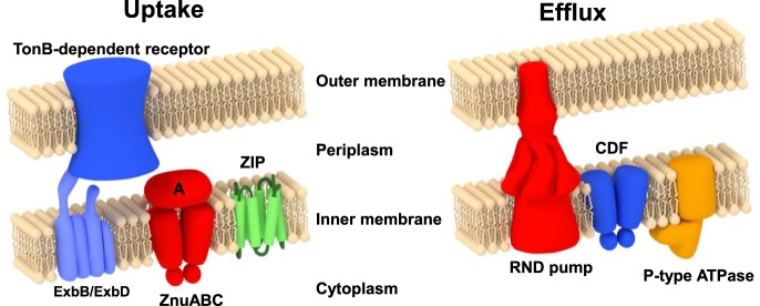
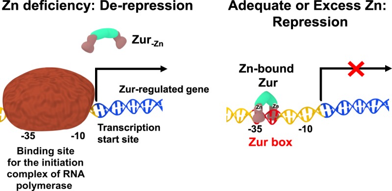
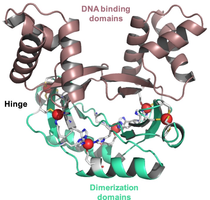
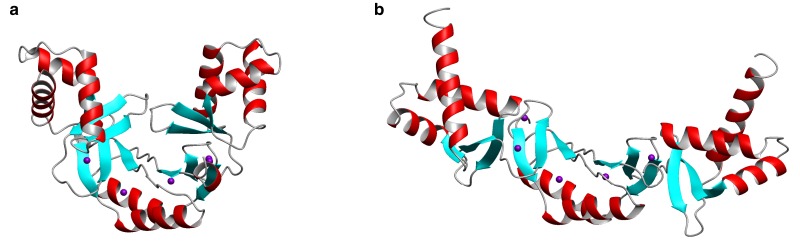
All organisms must regulate the cellular uptake, efflux, and intracellular trafficking of essential elements, including d-block metal ions. In bacteria, such regulation is achieved by the action of metal-responsive transcriptional regulators. Among several families of zinc-responsive transcription factors, the 'zinc uptake regulator' Zur is the most widespread. Zur normally represses transcription in its zinc-bound form, in which DNA-binding affinity is enhanced allosterically. Experimental and bioinformatic searches for Zur-regulated genes have revealed that in many cases, Zur proteins govern zinc homeostasis in a much more profound way than merely through the expression of uptake systems. Zur regulons also comprise biosynthetic clusters for metallophore synthesis, ribosomal proteins, enzymes, and virulence factors. In recognition of the importance of zinc homeostasis at the host-pathogen interface, studying Zur regulons of pathogenic bacteria is a particularly active current research area.

Keywords: Zur; bacteria; metal ions; zinc uptake regulator; zinc-responsive transcription factors.


Conflict of interest statement

The Authors declare that there are no competing interests associated with the manuscript.

Figures

Figure 1.
Figure 1.. Overview of the major players in bacterial zinc uptake and efflux, illustrated for a Gram-negative bacterium.
Proteins for import include members of the ZIP (zinc-iron permease) family and members of the ATP-binding cassette (ABC) superfamily. The latter systems consist of a membrane-bound permease, an ATPase, and a protein that is periplasmic in Gram-negative bacteria or on the cell surface in Gram-positive bacteria. These systems are usually named ZnuABC (Gram-negative bacteria) or AdcABC (Gram-positive bacteria), although this distinction is not consistently adhered to. A third label used frequently for such zinc importers is TroABC. Exporters include P-type ATPases, members of the cation–diffusion facilitator (CDF) family, and tripartite RND (root–nodulation–cell division) systems [3,10]. Regulatory proteins and further processes are explained in the main text.
Figure 2.
Figure 2.. Schematic illustration of canonical regulation of transcription by Zur.
Zinc-bound Zur (right-hand panel; see below for further details) represses transcription by binding to specific DNA sequences (Zur boxes) in the promoter region of Zur-regulated genes and thus inhibits initiation of transcription. When cells are deprived of zinc, demetallated Zur has a dramatically reduced affinity for DNA, allowing transcription to occur.
Figure 3.
Figure 3.. X-ray crystal structure of dimeric S. coelicolor Zur (pdb 3mwm) [38].
DNA-binding domains are shown in maroon and dimerisation domains in green. Zinc ions are shown as red spheres, nitrogen atoms in blue, sulfur in yellow, and carbon in light grey.
Figure 4.
Figure 4.. Conformational flexibility of Zur proteins.
The “closed” vs. “open” conformation of dimeric Zurs is illustrated by the X-ray structures of (A) ScZur (pdb 3mwm) and (B) MtZur (pdb 2o03).
Figure 5.
Figure 5.. Structural and sensory zinc sites on Zur proteins.
(A) ScZur (pdb 3mwm [38]) and (B) EcZur (pdb 4mtd [17]). The structural sites are highlighted in grey, the single or major sensory site in yellow, and the additional site in ScZur is highlighted in red. (C) Sequence alignment of Zur proteins from a variety of species. Residues confirmed to participate in zinc binding by X-ray crystallography are highlighted by red, yellow, and grey backgrounds. Residues involved in DNA binding are highlighted in cyan. Predicted metal-binding residues or sensory sites in Zur proteins that have not been structurally characterised are printed in red or yellow. The two residues forming a salt bridge in EcZur (see the text) are highlighted in green; they are (semi-)conserved in Zur from Salmonella, M. tuberculosis, and S. coelicolor.
Figure 6.
Figure 6.. DNA binding by EcZur.
(A) EcZur in complex with DNA (31 base pairs from the znuABC promoter; pdb 4mtd [17]). The two dimers binding to the complete Zur box are shown in green and purple. DNA backbone and bases are shown schematically, with the regions forming interactions with the protein highlighted in blue and magenta. The position of zinc ions is indicated in red. E. coli Zur boxes can bind one or two dimers; for the znuABC promoter, there is a high degree of cooperativity, leading to the overwhelming prevalence of the complex involving two dimers. (B) Illustration of the consensus sequence for the E. coli Zur box, with RNNNY (R = purine; Y = pyrimidine; N = any base) motifs important for Zur–DNA interactions highlighted. Each of the bars corresponds to the interaction motif for one monomer. The sequence logo for the consensus sequence is taken from ref. [17].
Figure 7.
Figure 7.. Examples for computationally assembled Zur boxes.
The sequence logos are taken directly from RegPrecise, using manually curated regulons [52]. While certain commonalities are evident for the Zur boxes from actinobacteria (Streptomycetaceae and Mycobacteriaceae) and Bacillales, the Zur boxes for enterobacteria (including E. coli and Salmonella) and cyanobacteria are clearly different.

References

    1. Dupont C.L. and Caetano-Anolles G. (2010) Mulkidjanian and Galperin: Zn may have constrained evolution during the Proterozoic but not the archean reply. Proc. Natl Acad. Sci. U.S.A. 107, E138 10.1073/pnas.1009565107 - DOI
    1. Andreini C., Banci L., Bertini I. and Rosato A. (2006) Zinc through the three domains of life. J. Proteome Res. 5, 3173–3178 10.1021/pr0603699 - DOI - PubMed
    1. Capdevila D.A., Wang J.F. and Giedroc D.P. (2016) Bacterial strategies to maintain zinc metallostasis at the host-pathogen interface. J. Biol. Chem. 291, 20858–20868 10.1074/jbc.R116.742023 - DOI - PMC - PubMed
    1. Outten C.E. and O'Halloran T.V. (2001) Femtomolar sensitivity of metalloregulatory proteins controlling zinc homeostasis. Science 292, 2488–2492 10.1126/science.1060331 - DOI - PubMed
    1. Neumann W., Gulati A. and Nolan E.M. (2017) Metal homeostasis in infectious disease: recent advances in bacterial metallophores and the human metal-withholding response. Curr. Opin. Chem. Biol. 37, 10–18 10.1016/j.cbpa.2016.09.012 - DOI - PMC - PubMed

Publication types

LinkOut - more resources