Review
. 2018 Aug 20;46(4):945-956.
doi: 10.1042/BST20170310. Epub 2018 Jul 31.

Protein motion in the nucleus: from anomalous diffusion to weak interactions

Affiliations
Review

Protein motion in the nucleus: from anomalous diffusion to weak interactions

Maxime Woringer et al. Biochem Soc Trans. .

Abstract

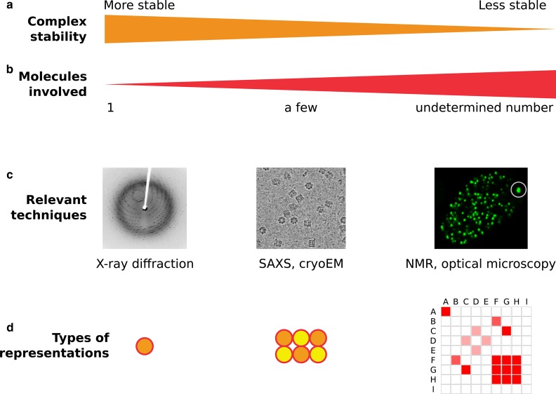
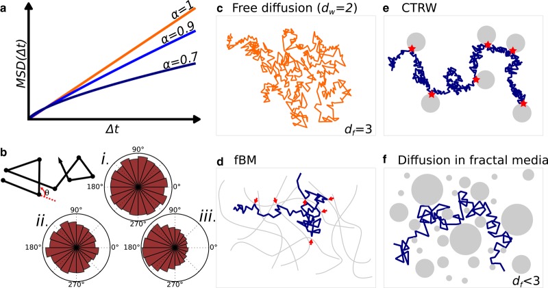
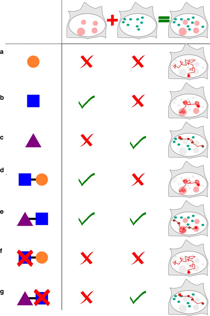
Understanding how transcription factors (TFs) regulate mammalian gene expression in space and time is a central topic in biology. To activate a gene, a TF has first to diffuse in the available space of the nucleus until it reaches a target DNA sequence or protein (target site). This eventually results in the recruitment of the whole transcriptional machinery. All these processes take place in the mammalian nucleoplasm, a highly organized and dynamic environment, in which some complexes transiently assemble and break apart, whereas others appear more stable. This diversity of dynamic behaviors arises from the number of biomolecules that make up the nucleoplasm and their pairwise interactions. Indeed, interactions energies that span several orders of magnitude, from covalent bounds to transient and dynamic interactions, can shape nuclear landscapes. Thus, the nuclear environment determines how frequently and how fast a TF contacts its target site, and it indirectly regulates gene expression. How exactly transient interactions are involved in the regulation of TF diffusion is unclear, but are reflected by live cell imaging techniques, including single-particle tracking (SPT). Overall, the macroscopic result of these microscopic interactions is almost always anomalous diffusion, a phenomenon widely studied and modeled. Here, we review the connections between the anomalous diffusion of a TF observed by SPT and the microscopic organization of the nucleus, including recently described topologically associated domains and dynamic phase-separated compartments. We propose that anomalous diffusion found in SPT data result from weak and transient interactions with dynamic nuclear substructures, and that SPT data analysis would benefit from a better description of such structures.

Keywords: molecular interactions; nuclear transport; protein motion; transcription factor.


Conflict of interest statement

The Author declare that there are no competing interests associated with this manuscript.

Figures

Figure 1.
Figure 1.. Diversity of weak interactions and example techniques to study them.
Biological interactions cover a wide spectrum in terms of complex stability (a) and number of molecules involved (b). This spectrum spans from stable protein complexes that can be purified and further imaged by techniques such as X-ray diffraction (left) to very labile, transient interactions that can involve thousands of proteins in vivo, whereas none of the interactions can be captured by traditional biochemistry (right). As one goes from one end of the spectrum to the other end, distinct sets of techniques (c) and types of representations (d) are needed. For instance, as the valency of interactions increases from a few strongly interacting partners to many weakly interacting partners, new graphical representations are needed, since traditional schematics representing macromolecular complexes whose stoichiometry is known as the juxtaposition of monomers (center) become difficult to read when depicting one protein weakly interacting with dozen of partners (right). In that case, matrices of pairwise interactions between proteins A-I might be more relevant. SAXS: Small-Angle X-Rays Scattering, NMR: Nuclear Magnetic Resonance.
Figure 2.
Figure 2.. Models of anomalous diffusion and plausible underlying physical structures.
(a and b) Characterizations of anomalous diffusion. (a) Sub-linear mean-square displacement plotted as a function of time characterizes subdiffusion, and reflects how a diffusing particle explores space, the degree of anomalous diffusion is characterized by the exponent α, the lower the α the more subdiffusive the process. (b) Free diffusion is characterized by isotropic distribution of angles — subpanel (i) — whereas an anisotropic distribution indicates anomalous diffusion — subpanels (ii) and (iii). (c) 3D free diffusion (dw=2), as usually encountered in a homogenous media (df=3). (df) Several types of heterogenous media can yield anomalous diffusion, including (d) diffusion within a viscoelastic polymer, in which a protein “bounces against” an elastic structure, a process traditionally described by fractional Brownian motion (fBm) and (e) free diffusion interspersed by long binding times — red stars, a process called CTRW, and (f) diffusion within a fractal media, that is a space obstructed by obstacles of all sizes.
Figure 3.
Figure 3.. Weak interactions of individual protein domains shape TF dynamics.
(a)–(c) Individual protein domains have specific transient interactions: the round domain (a) does not interact with any particular structure (represented by the two columns of the table), (b) the square domain interacts with a given pink structure (first column) and (c) the triangle domain interacts with the green structures (second column). This results in domain-specific dynamics (third column). (d and e) When protein domains are combined within a protein or TF, the observed SPT is a mixture between the interactions of each single domain. (f and g) When individual domains are mutated, the protein loses some transient specific interactions, and its dynamics can dramatically change (compare panels e and g).

References

    1. Sainsbury S., Bernecky C. and Cramer P. (2015) Structural basis of transcription initiation by RNA polymerase II. Nat. Rev. Mol. Cell Biol. 16, 129–143 10.1038/nrm3952 - DOI - PubMed
    1. Spitz F. (2016) Gene regulation at a distance: From remote enhancers to 3d regulatory ensembles. Semin. Cell Dev. Biol. 57, 57–67 10.1016/j.semcdb.2016.06.017 - DOI - PubMed
    1. Louder R.K., He Y., López-Blanco J.R., Fang J., Chacón P. and Nogales E. (2016) Structure of promoter-bound TFIID and model of human pre-initiation complex assembly. Nature 531, 604–609 10.1038/nature17394 - DOI - PMC - PubMed
    1. Murakami K., Tsai K.-L., Kalisman N., Bushnell D.A., Asturias F.J. and Kornberg R.D. (2015) Structure of an RNA polymerase II preinitiation complex. Proc. Natl Acad. Sci. U.S.A. 112, 13543–13548 10.1073/pnas.1518255112 - DOI - PMC - PubMed
    1. von Smoluchowski M. (1906) Zur kinetischen theorie der brownschen molekularbewegung und der suspensionen. Ann. Phys. 326, 756–780 10.1002/andp.19063261405 - DOI

Publication types