Review
. 2018 Aug 20;46(4):967-982.
doi: 10.1042/BST20170568. Epub 2018 Jul 31.

Emerging mechanisms of dynein transport in the cytoplasm versus the cilium

Affiliations
Review

Emerging mechanisms of dynein transport in the cytoplasm versus the cilium

Anthony J Roberts. Biochem Soc Trans. .

Abstract

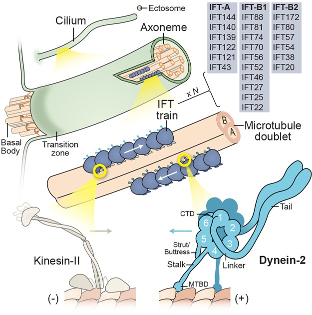
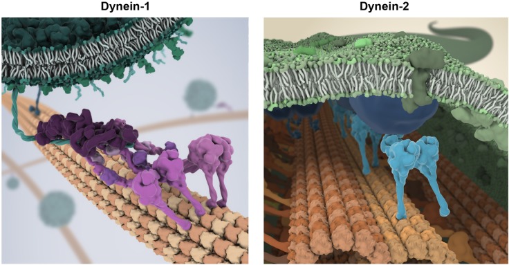
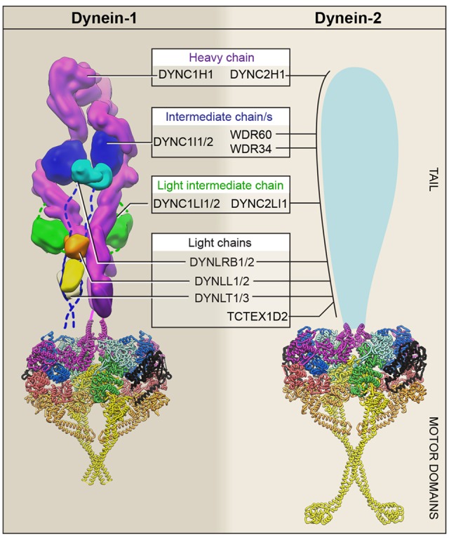
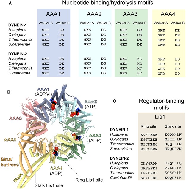
Two classes of dynein power long-distance cargo transport in different cellular contexts. Cytoplasmic dynein-1 is responsible for the majority of transport toward microtubule minus ends in the cell interior. Dynein-2, also known as intraflagellar transport dynein, moves cargoes along the axoneme of eukaryotic cilia and flagella. Both dyneins operate as large ATP-driven motor complexes, whose dysfunction is associated with a group of human disorders. But how similar are their mechanisms of action and regulation? To examine this question, this review focuses on recent advances in dynein-1 and -2 research, and probes to what extent the emerging principles of dynein-1 transport could apply to or differ from those of the less well-understood dynein-2 mechanoenzyme.

Keywords: cilia; dynein; intraflagellar transport; kinesin; microtubule.


Conflict of interest statement

Note added in proof

An analysis of dynein-2 subunit interactions via a visible immunoprecipitation (VIP) assay has been published, demonstrating interactions between WDR60 and TCTEX1/3-TCTEX1D2; WDR34 and LC8 and Roadblock; and the intermediate chain-light chain complex with the heavy chain and light-intermediate chain [148].

Figures

Box 1.
Box 1.. Cilia and intraflagellar transport.
Cilia fall into two broad classes: motile and non-motile. Motile cilia beat with a wave-like motion to either propel cells, such as sperm and protozoa, or generate flow over the cell surface. Conversely, a non-motile primary cilium is present on almost every cell type in the human body. A widespread view was that primary cilia represented nonfunctional vestigial structures. However, landmark discoveries recast primary cilia as the ‘signaling antenna’ of the cell [–151]. For example, mutations causing polycystic kidney disease were linked to an IFT subunit (IFT88) and a shortened cilia phenotype, supporting a sensory role for kidney primary cilia [152]. Hedgehog signaling, an important pathway for embryonic patterning, was found to depend on IFT proteins in mice [153] and involve dynamic localization of the receptor Smoothened to and from the ciliary membrane [154]. Other signaling components, involved in processes as diverse as sight, smell, taste, and appetite control, localize within cilia [150]. Moreover, receptor-containing ectosomes have recently been found to be secreted from the tip of the cilium to modulate signaling [–158]. The core of all cilia is the axoneme, a cylindrical array of typically nine microtubule doublets that extends from the basal body. Motile axonemes normally also have a central pair of microtubules, as well as periodic arrays of axonemal dyneins and regulatory complexes that co-ordinate ciliary beating [159]. The axoneme is covered by a ciliary membrane that is continuous with the plasma membrane but distinct in protein and lipid content. During ciliary growth and maintenance, new subunits are incorporated at the ciliary tip, where the microtubule plus ends (+) reside. A diffusion barrier separates the ciliary volume and the cytoplasm [160,161]. This selective barrier involves the ‘transition zone’, a region immediately distal to the basal body characterized by Y-shaped links between the doublets and the ciliary membrane. Anterograde IFT, powered by kinesin-II motors, moves cargoes synthesized in the cytoplasm through the transition zone and toward the tip of the cilium. Conversely, dynein-2 returns cargoes to the cell body. Both motors associate with IFT trains, polymeric arrays involving two sub-complexes, IFT-A and IFT-B (consisting of IFT-B1 and IFT-B2) [162,163]. Genetically, IFT-B proteins tend to be critical for anterograde IFT and ciliogenesis, while IFT-A proteins are typically linked with retrograde IFT. However, the functions of IFT-A and -B are not so simply separated, as IFT-B proteins can be involved in cargo export, while IFT-A proteins are required for ciliary entry of a subset of membrane proteins via the adaptor protein TULP3 [164]. Structurally, dynein-2 comprises a tail domain and a motor domain containing the linker, a ring of six AAA+ modules (1–6), a coiled-coil stalk with the MTBD at its tip, a shorter coiled-coil strut/buttress, and a C-terminal domain (CTD).
Figure 1.
Figure 1.. Speculative impressions of cargo transport by dynein-1 and dynein-2.
Left: Depiction of dynein-1 transporting a vesicle. Two dynein-1 complexes (magenta) are templated by dynactin (dark purple) and a coiled-coil cargo adaptor (teal), whose distal end attaches to a receptor on the vesicle surface. For clarity, the densely packed milieu of molecules in the cytoplasm is not shown. Right: Depiction of dynein-2 (cyan) propeling a retrograde IFT train (blue) within the cilium. Dynein-2 operates in the confined space between the ciliary membrane (green) and the axoneme (orange), moving on the A-tubule of the microtubule doublet. Artwork in collaboration with Bara Krautz (www.scienceanimated.com; email: bara@scienceanimated.com).
Figure 2.
Figure 2.. Dynein-1 and -2 subunit composition.
Summary of unique and shared components in dynein-1 and -2. The C-terminal region of each heavy chain forms the motor domain, while the N-terminal region forms the tail and associates with intermediate, light-intermediate, and light chains. In mammals, there are two isoforms for each class of dynein-1-associated subunit. For example, the two intermediate chain isoforms as denoted here as ‘DYNC1I1/2’. Structural information is available for the dynein-1 and -2 motor domains, which are shown in their auto-inhibited ‘phi-particle’ state in ribbon representation (colored as in Figure 3) [57,92,126]. The dynein-1 tail is shown in surface representation [57]. The unknown architecture of the dynein-2 tail is shown schematically.
Figure 3.
Figure 3.. Dynein motor domain structure and motif conservation.
(A) Analysis of the Walker-A and -B motifs within the AAA+ modules of dynein-1 and -2 from different species. Within the Walker-A consensus sequence (GKT), the lysine is important for nucleotide binding. Within the Walker-B consensus sequence (DE), the glutamic acid is thought to be the catalytic base that polarizes H2O for an inline attack on the γ-phosphate of ATP. Amino acids matching the consensus sequence are shown in bold type. In dynein-2, the Walker-A and -B motifs of AAA3 and AAA4 deviate from the consensus, in contrast with the situation in dynein-1. Beyond the AAA+ modules, Redwine et al. [165] have noted differences in charged amino acids in the MTBD of dynein-1 and dynein-2, which are likely to influence their respective microtubule affinities. (B) Structure of the dynein-2 AAA+ ring from PDB 4RH7 [92], with α-helices shown as cylinders and nucleotides in space-filling representation. The linker domain, which would lie on the near face of the AAA+ ring, and the CTD, which would lie on the far face, are omitted for clarity. (C) Analysis of Lis1's binding sites in the AAA+ ring and stalk. Key charged and polar amino acids (bold) shown to be important for Lis1 binding in S. cerevisiae dynein-1 [108,110] are not conserved in dynein-2.

References

    1. Wickstead B. and Gull K. (2007) Dyneins across eukaryotes: a comparative genomic analysis. Traffic 8, 1708–1721 10.1111/j.1600-0854.2007.00646.x - DOI - PMC - PubMed
    1. Kollmar M. (2016) Fine-tuning motile cilia and flagella: evolution of the dynein motor proteins from plants to humans at high resolution. Mol. Biol. Evol. 33, 3249–3267 10.1093/molbev/msw213 - DOI - PMC - PubMed
    1. Wilkes D.E., Watson H.E., Mitchell D.R. and Asai D.J. (2008) Twenty-five dyneins in Tetrahymena: a re-examination of the multidynein hypothesis. Cell Motil. Cytoskeleton 65, 342–351 10.1002/cm.20264 - DOI - PubMed
    1. Yagi T. (2009) Bioinformatic approaches to dynein heavy chain classification. Methods Cell Biol. 92, 1–9 10.1016/S0091-679X(08)92001-X - DOI - PubMed
    1. Paschal B.M., Shpetner H.S. and Vallee R.B. (1987) MAP 1c is a microtubule-activated ATPase which translocates microtubules in vitro and has dynein-like properties. J. Cell Biol. 105, 1273–1282 10.1083/jcb.105.3.1273 - DOI - PMC - PubMed

Publication types

LinkOut - more resources