

Review
doi: 10.1016/j.jmu.2017.08.002.
Epub 2017 Sep 12.
Total Anomalous Pulmonary Venous Connection: From Embryology to a Prenatal Ultrasound Diagnostic Update
Affiliations
- PMID: 30065477
- PMCID: PMC6029298
- DOI: 10.1016/j.jmu.2017.08.002
Item in Clipboard
Review
Total Anomalous Pulmonary Venous Connection: From Embryology to a Prenatal Ultrasound Diagnostic Update
J Med Ultrasound.
2017 Jul-Sep.
No abstract available
Keywords: Fetal venous system; Prenatal ultrasound; TAPVC; Total anomalous pulmonary connection.
Conflict of interest statement
Conflict of interest: The authors have no conflicts of interest relevant to this article.
Figures
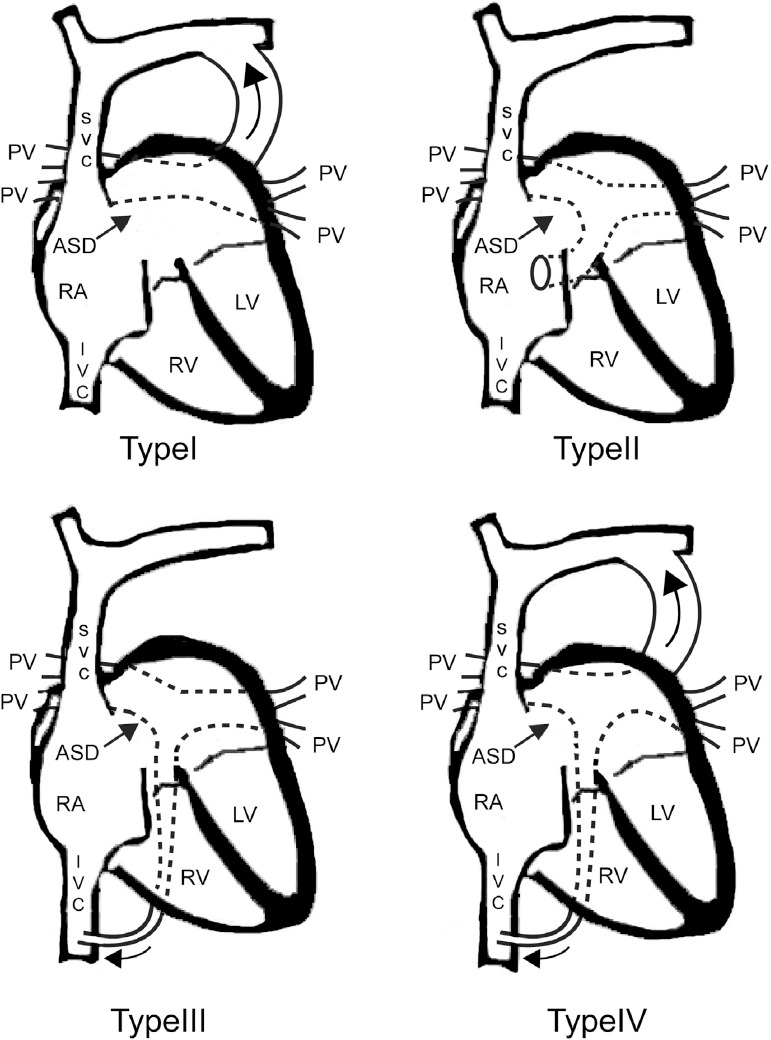
Darling classification of total anomalous pulmonary venous connection (TAPVC). Type I: supracardiac type. Four pulmonary veins connect to a vertical vein which subsequently drains into the left brachiocephalic vein and superior vena cava (curved arrow). Type II: intracardiac type. The pulmonary veins empty into the coronary sinus or directly into the right atrium. Type III: infracardiac type. The pulmonary veins connect to a vertical vein passes through diaphragm. The pulmonary flow ultimately drains to the systemic venous system, such as portal vein or inferior vena cava (curved arrow). Type IV: mixed type. ASD, atrial septum defect; LV, left antrum; IVC, inferior vena cava; PV, pulmonary vein; RA, right antrum; SVC, superior vena cava.

On an apical four-chamber view, right and left inferior pulmonary vein enters left antrum posteriorly and forms a “horn-like” insertion. Note the posterior wall of left antrum is not as round and smooth as right antrum.

Four-chamber view of the right pulmonary vein empty into left antrum in color Doppler image. Note the color box should be narrowed to the LA with low pulse-repetition frequency (0.8–2.0 Hz) and high sensitivity setting.

Doppler waveform across the inferior pulmonary vein. This triphasic pattern is similar to the waveform observed in the ductus venosus. Note the Doppler sample gate placed on the pulmonary vein is within the lung parenchyma (s, systolic velocity; d, diastolic velocity; a, atrial reversal flow).
References
-
- Hoffman JI, Kaplan S. The incidence of congenital heart disease. J Am Coll Cardiol. 2002 Jun 19;39(12):1890–900. Review. - PubMed
-
- Seale AN, Uemura H, Webber SA, et al. British Congenital Cardiac Association. Total anomalous pulmonary venous connection: morphology and outcome from an international population-based study. Circulation. 2010 Dec 21;122(25):2718–26. - PubMed
-
- Hoffman JI, Kaplan S, Liberthson RR. Prevalence of congenital heart disease. Am Heart J. 2004 Mar;147(3):425–39. - PubMed
-
- Wu MH, Chen HC, Lu CW, et al. Prevalence of congenital heart disease at live birth in Taiwan. J Pediatr. 2010 May;156(5):782–5. - PubMed
Publication types
LinkOut - more resources
Full Text Sources
Other Literature Sources