Rare Brain Tumor in a Neonate
- PMID: 30065500
- PMCID: PMC6029326
- DOI: 10.1016/j.jmu.2017.09.004
Rare Brain Tumor in a Neonate
Abstract
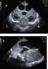
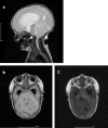
Neonatal brain tumor is rare and its outcome is generally poor. We reported a 17-day-old neonate presented as enlarged head girth. The pathological finding showed an embryonal tumor with multilayered rosettes.
Keywords: Brain tumors; ETMR; Neonate; Ultrasonography.
Conflict of interest statement
Conflicts of interest: There are no conflicts of interest.
Figures


References
-
- Buetow PC, Smirniotopoulos JG, Done S. Congenital brain tumors: a review of 45 cases. AJR Am J Roentgenol. 1990;155(3):587–93. - PubMed
-
- Johnson ML, Mack LA, Rumack CM, et al. B-mode echoence-phalography in the normal and high risk infant. Am J Roent-genol. 1979;133(3):375–81. - PubMed
-
- Isaacs H. Perinatal brain tumors: a review of 250 cases. Pediatr Neurol. 2002;27:249–61. - PubMed
-
- Magdum SA. Neonatal brain tumours - a review. Early Hum Dev. 2010;86(10):627–31. - PubMed
-
- Paulus W, Kleihues P. Genetic profiling of CNS tumors extends histological classification. Acta Neuropathol. 2010;120(2):269–70. - PubMed
Publication types
LinkOut - more resources
Full Text Sources
Other Literature Sources
